We are back to welcome the new year as we love to do with the Atlantic Coast Retina Club (ACRC). This legendary regional meeting is now over 40 years in the running. ACRC has rotated between Baltimore, New York and Philadelphia, and more recently Boston as well. The 2025 ACRC was held in Baltimore at the Wilmer Eye Institute / Johns Hopkins University. ACRC runs for 2 days: the first day is comprised of mystery cases presented by trainees, and the second day is by faculty. The third day is the Macula meeting, where faculty from throughout the Atlantic Coast provide updates on timely topics. We hope you enjoy the RETINA Roundup coverage of the ACRC and Macula 2025 meetings.
RETINA Roundup Editorial Team
Saif Hamdan, Wills Eye Hospital
Vishal Swaminathan, Mayo Clinic
SESSION #1
The second day of the Atlantic Coast Retina Club was kicked off with another series of mystery cases presented by trainees and moderated by Drs. Adrienne Scott and John Thompson.
First, Dr. Lisa Koeinig (Memorial Sloan Kettering Cancer Center) shared a case of a young girl with unilateral vision loss, highlighting several important diagnostic and management considerations for ocular manifestations of Sturge-Weber syndrome (SWS) including diffuse choroidal hemangioma. Interestingly, the patient did not feature a port-wine stain at time of presentation, though as noted by Dr. Carol Shields, these may have been treated with pulse dye laser therapy or rarely can undergo spontaneous regression.
In the next talk, Dr. Ryan (Sameen) Meshkin of Mass Eye and Ear shared a case of a woman in her 30s with stage 3c low-grade serous peritoneal carcinoma on chemotherapy presenting with one month of bilateral blurry vision. On further testing, the patient had multifocal peripheral vascular leakage in areas of nonperfusion on fluorescein angiography, but interestingly without macular or disc leakage. She also had bulls-eye hyper-autofluorescence peripherally and scattered areas of hyper- and hypo-autofluorescence extending nasally on fundus autofluorescence testing, clinching a diagnosis of angiographically silent cystoid macular edema (CME) secondary to paclitaxel toxicity, compounded by underlying Enhanced S-Cone Syndrome (ESCS). The diagnosis was further supported by genetic testing showing pathogenic homozygous mutation in the NR2E3 gene and a history of lifelong night vision difficulties in the patient’s sibling. Upon discontinuation of paclitaxel, the CME resolved in the left eye but persisted in the right eye and continues to be managed. This unique case highlights the interplay between inherited retinal disease (ESCS) and chemotoxicity (paclitaxel-induced CME).

Next, Dr. Isaac Bleicher (Mass Eye and Ear) revealed a fascinating example of a patient with VKH-like reaction secondary to DRESS syndrome, highlighting the complexity of autoimmune and inflammatory responses triggered by hypersensitivity reactions. It was noted by several faculty that similar VKH-like reactions have also been seen with checkpoint inhibitors.

Dr. Taweevat Attaseth from the Wills Eye Hospital Ocular Oncology service was the next speaker, describing an atypical presentation of multiple choroidal melanomas in the same eye of a man in his 60s. The case highlighted oculodermal melanocytosis as a known risk factor for malignant transformation into melanoma, especially with multifocal lesions and the need for lifelong monitoring. It emphasized how FAF and ultrasound play a pivotal role in detecting small, multifocal lesions and assessing progression. There were interesting discussions among the audience and moderators regarding accurate measurement of tumor thickness when utilizing A- and B-scan.

SESSION #2
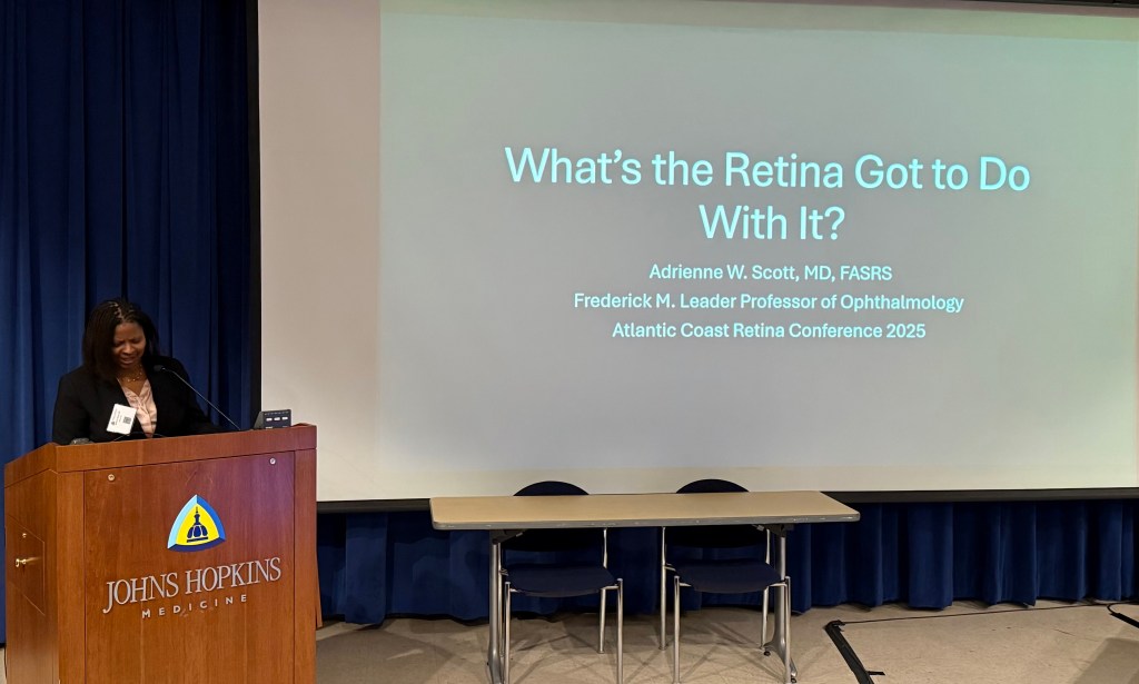
The following session featuring case presentations by faculty was moderated by Drs. Bailey Freund and Sunir Garg. The first case by Dr. Adrienne Scott of Wilmer shared her case entitled “What’s the Retina Got to Do With It?” discussing a young child admitted with acute unilateral periorbital swelling and anemia found to have acute orbital bone infarction, a rare complication of sickle cell disease. She stressed the importance of early outpatient surveillance in these children even in the absence of visual symptoms, stressing early features of sickle cell retinopathy such as temporal macular thinning on OCT.

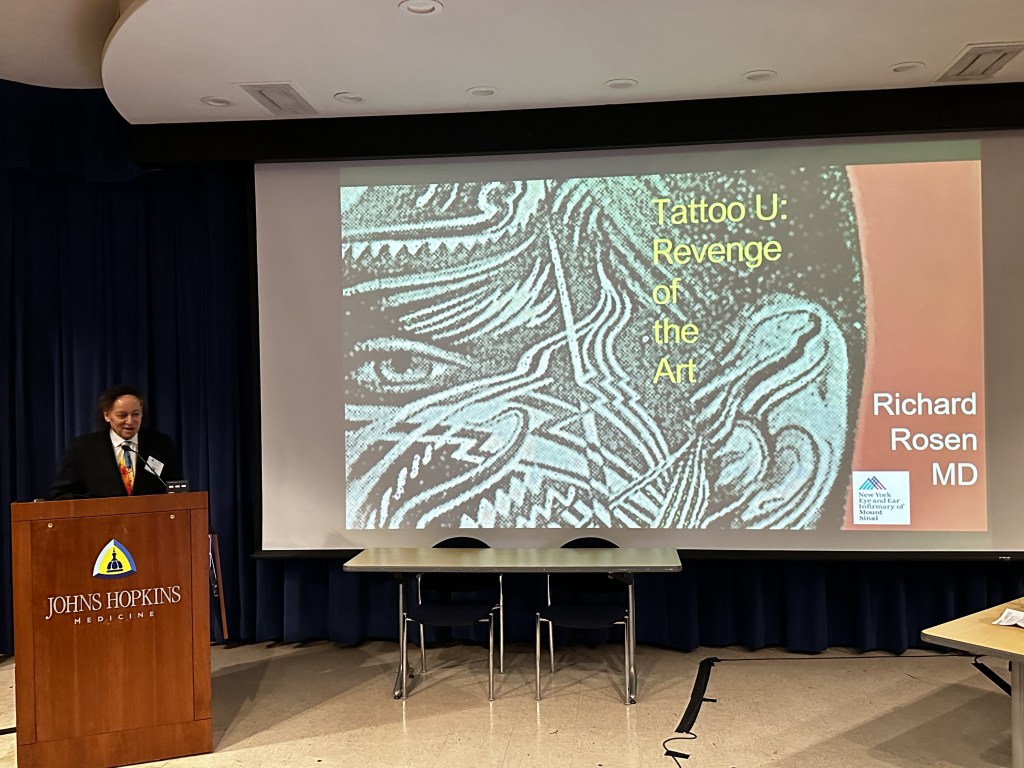
Next, Dr. Richard Rosen of New York Eye and Ear presented an interesting case of a tattoo removal practitioner with laser-induced retinal injury associated with occupational exposure during tattoo removal. She had known exposure to 1064 nm lasers used for tattoo removal. Fundus exam was notable for subretinal hemorrhage near the macula with possible early macular hole, and fundus autofluorescence imaging showed blockage from hemorrhage and possible window defects exposing the RPE. OCT showed evidence of retinal thinning. This case emphasized how laser retinal injuries are underreported, especially in cosmetic fields where high-powered devices are used, and the importance of education regarding this topic and stricter occupational guidelines.

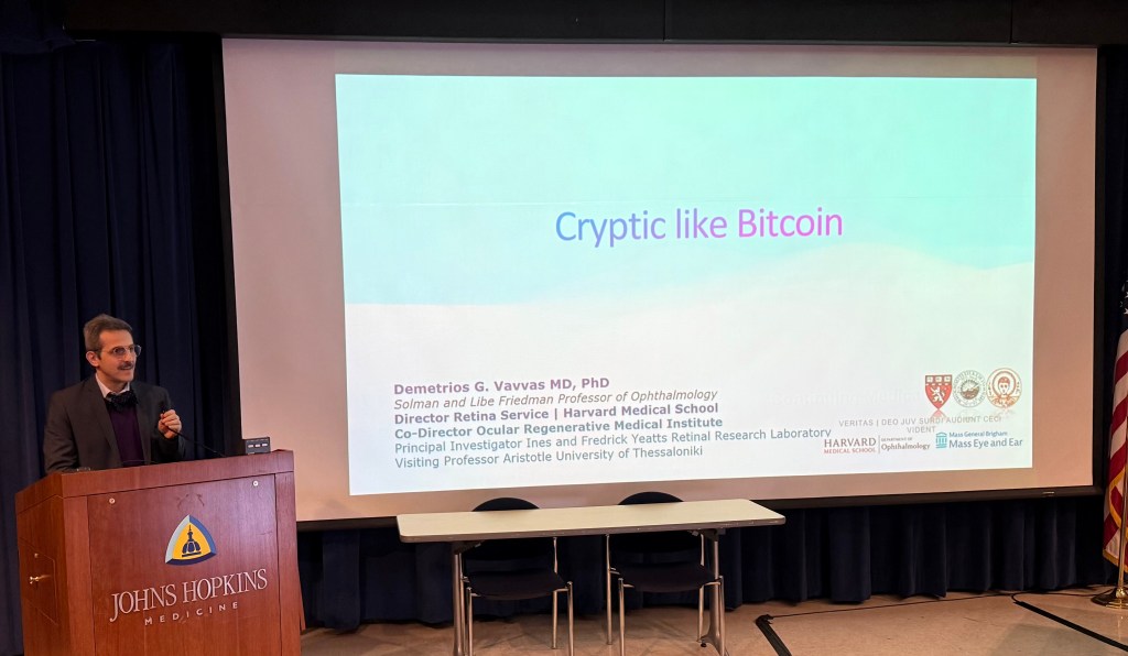
Dr. Demetrios Vavvas of Mass Eye and Ear then shared an atypical case presentation of cryptococcus retinitis (rather than the classical choroiditis) in an immunosuppressed patient. He highlighted how infectious etiologies such as cryptococcus and toxoplasmosis often present and act differently in the immunocompromised host. The moderators commented that the title of his talk gave away the diagnosis very quickly!

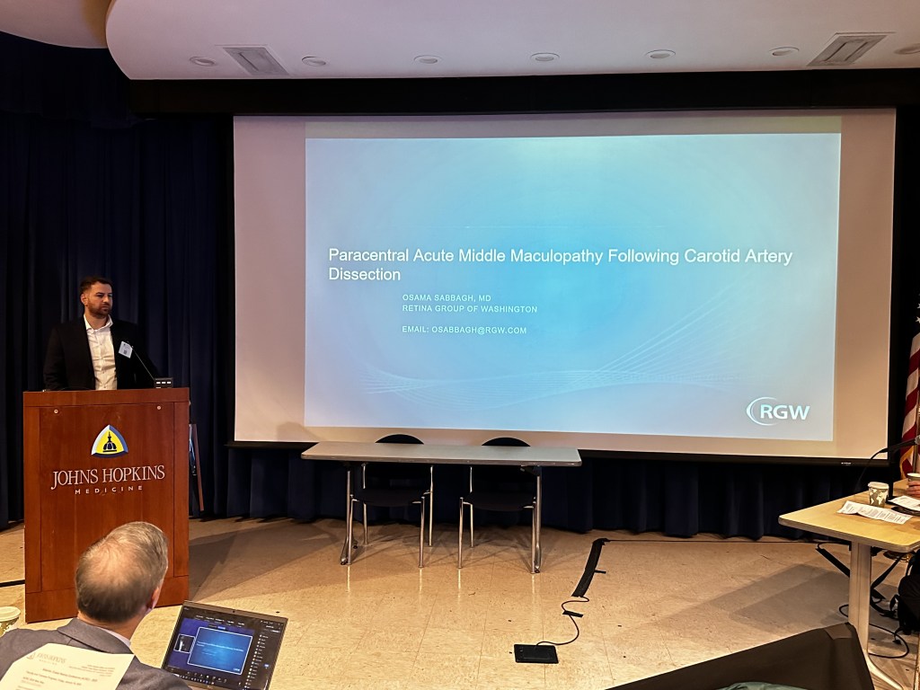
Next, Osama Sabbagh of the Retina Group of Washington described a woman in her 30s presenting with multiple episodes of transient vision loss with residual scotoma in the left eye, along with numerous systemic symptoms such as paresthesias and dysarthria. She was found to have paracentral acute middle maculopathy (PAMM) following a carotid artery dissection. PAMM represents ischemia at the level of the deep retinal capillary plexus and in this interesting case, it was likely secondary to slow or impaired perfusion caused by arterial insufficiency from the carotid artery dissection.

Lastly, Dr. Nauman Chaudhry from Retina Group of New England discussed a case series of patients undergoing treatment with complement inhibitors for geographic atrophy that developed macular neovascularization (MNV). While a well-known complication, he highlights the importance of thorough screening to assess for active or subclinical MNV before initiating complement inhibitors as well as close monitoring.

Read All Atlantic Coast Retina Club / Macula 2025 Articles:
Mystery Cases 1 & 2
Mystery Cases 3 & 4
Mystery Cases 5 & 6
Mystery Cases 7, 8, 9
Imaging, GA, and IRDs
Keynote Lectures
DR and DME
Choroidal Neovascularization
Retinal Vascular Diseases
Ocular Oncology
Special Topics & Women in Retina