Roselind Ni and Bita Momenaei
Wills Eye Hospital
SESSION #3
The third morning session, moderated by Dean Eliott, MD and William Mieler, MD, featured four compelling case presentations, sparking insightful discussions among the attending retina specialists.
Dr. Thamolwan Surakiatchanukul from the Geisel School of Medicine at Dartmouth, first presented a case of a an elderly woman with floaters in her right eye, significant anterior chamber and vitreous inflammation, and areas of retinal whitening with intralesional hemorrhages. The left eye showed anterior chamber cells but no fundus abnormalities. An AC tap confirmed varicella-zoster virus (VZV) as the cause, leading to a diagnosis of acute retinal necrosis (ARN) due to VZV. The patient was treated with intravitreal foscarnet, high-dose systemic valacyclovir, and Pred Forte.
ARN is typically associated with VZV in older adults and HSV-2 in younger patients, most commonly presenting in immunocompetent individuals, though it occurs in about 15% of immunocompromised cases. Approximately 30% of cases may exhibit retinal hemorrhages. The contralateral eye is at the highest risk of involvement within 14 weeks, but systemic antiviral therapy significantly reduces this risk. ARN has an RD risk of up to 85%, making lesion size and activity control crucial through aggressive antiviral therapy. The case fostered a dynamic discussion, emphasizing key management strategies:
- Role of Laser Therapy: Dr. Arevalo noted that laser barricades are ineffective in preventing RD progression but highlighted the importance of combining intravitreal antivirals with steroids (after 48 hours to avoid disease exacerbation).
- Prophylactic Vitrectomy: Dr. Arevalo and Dr. Rosen emphasized its benefits in patients with persistent or fovea-threatening lesions, noting its ability to remove inflammatory cytokines and improve outcomes.
- Intravitreal Foscarnet: Frequently used to manage ARN, with Dr. Rosen highlighting the importance of monitoring for increased vitreous inflammation post-treatment and intervening early with vitrectomy if needed.
- Duration of Prophylactic Treatment: Dr. Arevalo suggested indefinite systemic antiviral prophylaxis when feasible, although no consensus exists.
Dr. Yoshihiro Yonekawa from Wills Eye Hospital next shared a rare and intriguing case of a one-year-old child initially diagnosed with bilateral retinal detachments since birth. Examination under anesthesia revealed bullous retina positioned directly behind the lens, with yellowish subretinal exudates in both eyes. The anterior segments appeared normal, and ultrasound imaging showed no masses. Genetic testing confirmed a severe type of X-linked retinoschisis caused by an RS1 mutation.
Dr. Yonekawa proposed three potential approaches to the audience, sparking a lively debate about the risks and benefits of each option:
- External drainage and creating only an outer retinal break.
- Vitrectomy with a small retinotomy and oil tamponade.
- Scleral buckle combined with vitrectomy and oil tamponade
Dr. Eliott recounted a case involving macular schisis with vitreous hemorrhage, where internal limiting membrane peeling improved visual outcomes. Dr. Jampol proposed the use of Diamox in select cases.
Dr. Yonekawa finally recommended “none of the above!” noting, “sails can furl on their own.”
He shared follow-up findings at six and twelve months, where both eyes showed spontaneous resolution of the schisis cavities. While not all cases resolve spontaneously, this outcome underscores the importance of careful observation before pursuing high-risk surgical interventions.

Dr. K. Bailey Freund from NYU Grossman School of Medicine presented the case of an elderly woman with age-related macular degeneration (AMD) and early macular atrophy, highlighting the challenges of visualizing atrophy margins using various autofluorescence imaging modalities. While OCT confirmed macular atrophy, the Optos device struggled to delineate margins, particularly with green autofluorescence. Blue autofluorescence on the EIDON camera provided clearer visualization, though it absorbed centrally due to luteal pigment, creating a dark area.
Dr. Freund noted that in cases with a choroidal atrophy, the collagen’s autofluorescence becomes visible. This is significant because these patients often experience notable visual symptoms before macular atrophy becomes apparent. He emphasized that this issue is not exclusive to the Optos; other imaging systems also face challenges in distinguishing the borders of atrophy in high myopes with lepto-choroid.
He discussed how device-specific factors like wavelength, confocality, and averaging influence imaging outcomes. Devices like the SPECTRALIS, with high confocality, excel in delineating atrophic areas, while the Optos, with lower confocality, offers depth of field but less precision for atrophy borders.

Dr. Jay Chhablani from the University of Pittsburgh shared the case of a highly myopic woman who presented with sudden-onset vision loss in the right eye. Initial imaging showed subretinal fluid and leakage on fluorescein angiography, leading to a presumed diagnosis of central serous chorioretinopathy (CSCR). Follow-up imaging 1.5 months later revealed subretinal hemorrhage, and further evaluation diagnosed choroidal neovascularization (CNV).
A key observation was localized choroidal thickening with atypically fuzzy vessels, rather than diffuse changes. OCTA indicated CNV, and detailed angiography revealed multiple small leakages instead of a single ink-blot pattern. These findings pointed to punctate inner choroidopathy (PIC) as the underlying cause, with the localized thickening aligning with the characteristic “sponge sign” of PIC. The patient was diagnosed with CNV secondary to PIC and treated with one intravitreal anti-VEGF injection, leading to significant improvement. Following a second injection, she achieved 20/20 vision and remained asymptomatic at her one-year follow-up without requiring further treatment. The case prompted an engaging discussion focused on management options:
- Steroid Use in Inflammatory Conditions accompanied by CSCR:
In response to Dr. Arevalo’s question about systemic steroids in inflammatory conditions like multifocal choroiditis, Dr. Chhablani explained that not every patient with CSCR responds poorly to steroids, nor does every steroid exacerbate CSCR. Systemic immunosuppressants or oral steroids are preferred when inflammation is the primary driver. Oral steroids are favored as they allow for titration and tapering to monitor response and recurrence. However, he cautioned that local steroids might exacerbate CSCR. - Importance of Early OCTA:
Dr. Rosen highlighted the value of early OCTA in detecting neovascularization that may be missed on fluorescein angiography, emphasizing its utility in ambiguous presentations.

SESSION #4
The fourth session of the day was moderated by Carl Regillo, MD, and Richard Rosen, MD.
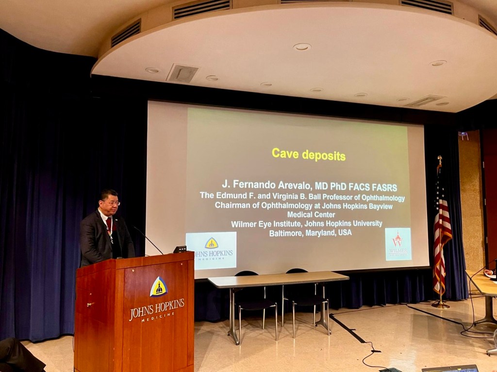
This session started off with Dr. Fernando Arevalo from Wilmer Eye Institute and co-chair of the meeting, presenting a case of toxoplasmosis gondii infection. The patient presented with preretinal inflammatory deposits, stalactite-like deposits on the surface of the retina seen on OCT, along with peripheral peripheral necrotizing retinitis. The case emphasized that these deposits should not be overlooked, and are a marker of active disease, indicating an infectious or inflammatory condition. PCR is important for the diagnosis, and the infection was treated with bactrim and prednisone.

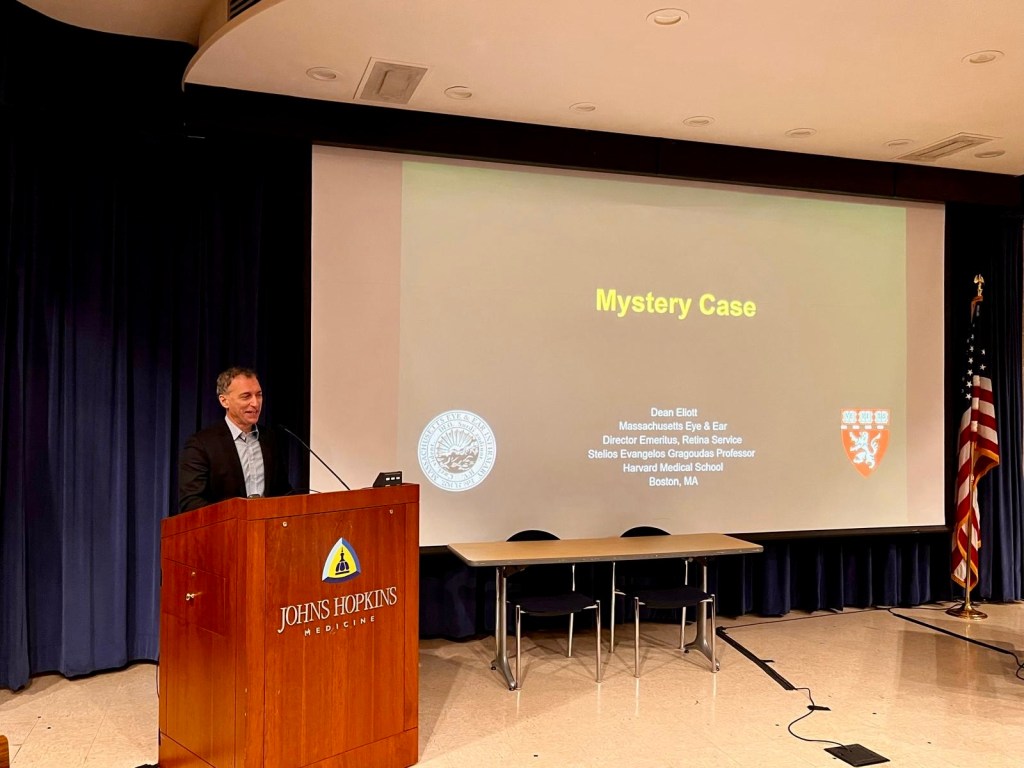
Next, Dr. Dean Elliot from Mass Eye & Ear presented a case of drug-induced helochoid peripapillary chorioretinal dystrophy that mimicked Sveinsson’s Chorioretinal Atrophy. This is a rare, autosomal dominant condition caused by a mutation in TEA domain transcription factor 1 (TEAD1), which plays a role in cellular proliferation, and may be targeted in cancer treatements. The patient had presented with blurry vision, and peripapillary radial hypopigmentation. They were found to have been taking a YAP/TEAD1 inhibitor for treatment of mesothelioma about 8 to 4 months before presentation before changing their drug regimen, and this was believed to be the cause for her findings, due to the underlying common pathogenesis of Sveinsson’s Chorioretinal Atrophy and the pharmacologic mechanism of TEAD1 inhibitors.

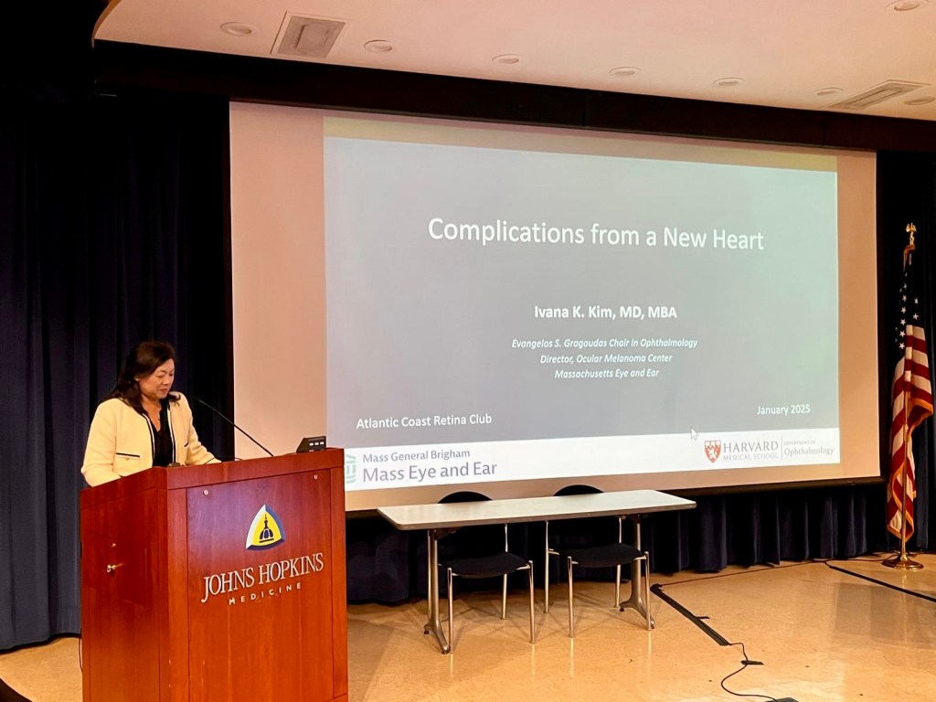
To continue, Dr. Ivana Kim, also of Mass Eye & Ear, presented “Complications from a New Heart”, which was a case of endogenous Nocardia endophthalmitis and disseminated nocardiosis in a heart transplant patient. The patient was on immunosuppresive therapy, and was in the ICU for weakness and dyspnea. They presented with blurry vision, imaging showed brain enhancing lesions and pulmonary lesions, and bronchoscopy and blood culture confirmed Nocardia. The patient had a prolonged ocular course – they received more than a year of intraocular injections and systemic antibiotics, balancing efficacy and toxicity of different drugs, and vitrectomy without debridement of the subretinal abscess. Ultimately the systemic and ocular infection was resolved with visual acuity at 20/600 and 20/25 two years after presentation.

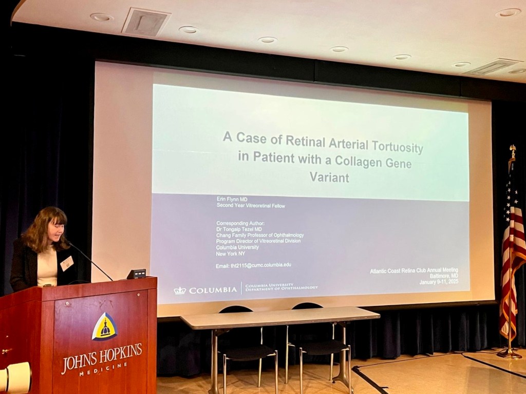
Finally, Dr. Erin Flynn, a second year vitreoretinal fellow at Columbia, presented “A Case of Retinal Arterial Tortuosity in Patient with a Collagen Gene Variant”. The patient, a teenage woman, presented with floaters, and marked retinal arterial tortuosity starting from the optic nerve to the periphery. The patient’s father was seen as well and also had retinal arterial tortuosity. Genetic testing was done for both patient and father, and they were found to have a novel mutation in COL1A1, causing a defect in the type 1 collagen alpha-1 chain. The geneticist ruled out classic and hypermobility Ehler’s Danlos syndrome, however Dr. Flynn brought up that there is a subtype associated with type 1 collagen mutations which has an overlap with osteogenesis imperfecta and vascular abnormalities in the gastrointestinal system. Dr. Pulido suggested that with tortuosity starting at the disc, patients should be worked up for Fabry syndrome. Dr. Freund commented on the case’s similarities with familial retinal arterial tortuosity, which is a collagen type 4 defect, with tortuosity beginning after the second or third bifurcation. The importance of checking the patient’s risk for stroke when the patient first presents was also emphasized.

Read All Atlantic Coast Retina Club / Macula 2025 Articles:
Mystery Cases 1 & 2
Mystery Cases 3 & 4
Mystery Cases 5 & 6
Mystery Cases 7, 8, 9
Imaging, GA, and IRDs
Keynote Lectures
DR and DME
Choroidal Neovascularization
Retinal Vascular Diseases
Ocular Oncology
Special Topics & Women in Retina