Yasman Moshiri, MD
Bascom Palmer Eye Institute, Miami, FL
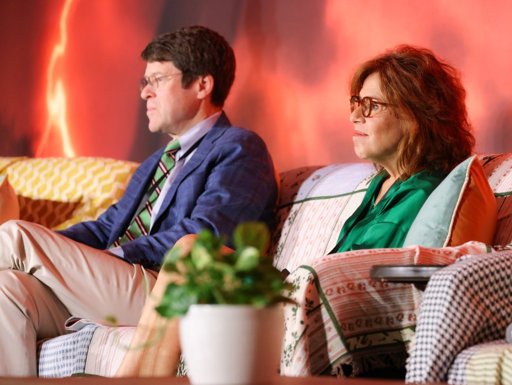
The first session of the 12th Annual Meeting of the Vit-Buckle Society was an exciting foray into the clinical and surgical decision-making of some of the most seasoned retina specialists. The session was moderated by Dr. Maria Berrocal and Dr. Baker Hubbard.
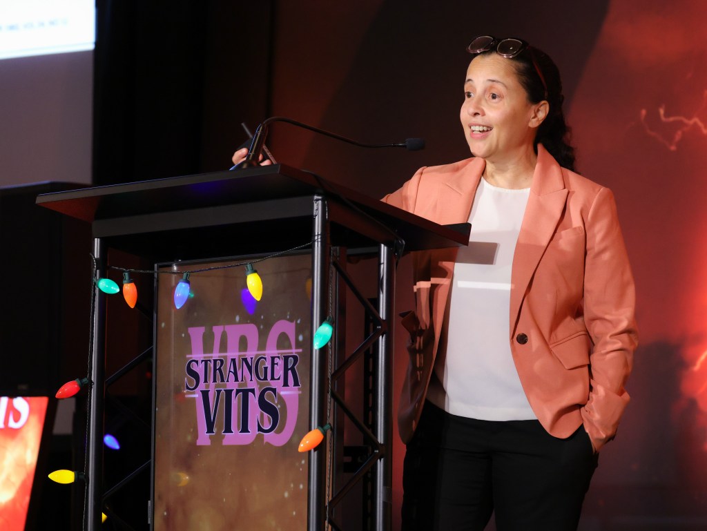
The session began with an in-depth discussion on Surgery for Diabetic Retinopathy: Pearls and Pitfalls by Dr. Amani A. Fawzi, MD, Cyrus Tang and Lee Jampol Professor of Ophthalmology at Northwestern University. In her talk, Dr. Fawzi outlines several clinical pearls to optimize outcomes in TRD surgery. She begins with “Respect the Hyaloid!” Determine whether the hyaloid has separated. One preoperative clue for this is whether the patient has undergone PRP. If it’s unclear, OCT can help. Dr. Fawzi goes on to say, “Think about where it’s attached, where the plaques are, where you’re going to lift, and how you’re going to segment.” Intraoperatively, Dr. Fawzi recommends staining the hyaloid, staining again, and staining again even when you think you’re done – and be careful around the vessels! She states that if there is a fibrosed Weiss ring visible above the nerve, start there using forceps. This opens the subhyaloid space for access. Another key pearl she shares is to leave the nasal retina until the end of the TRD surgery, because this is where the hyaloid is most attached and there is risk for iatrogenic tears. She reminds the audience to maximize preoperative PRP if the TRD is not involving the macula, especially in type 1 diabetics, as they may be prone to fibrinoid reactions.

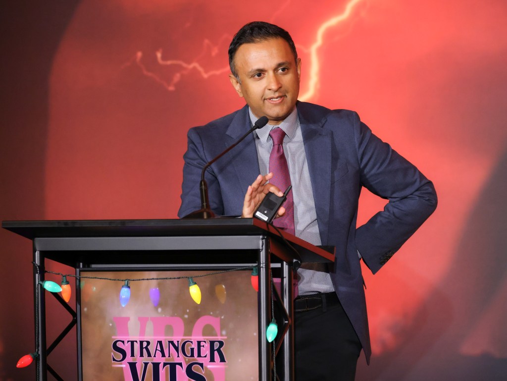
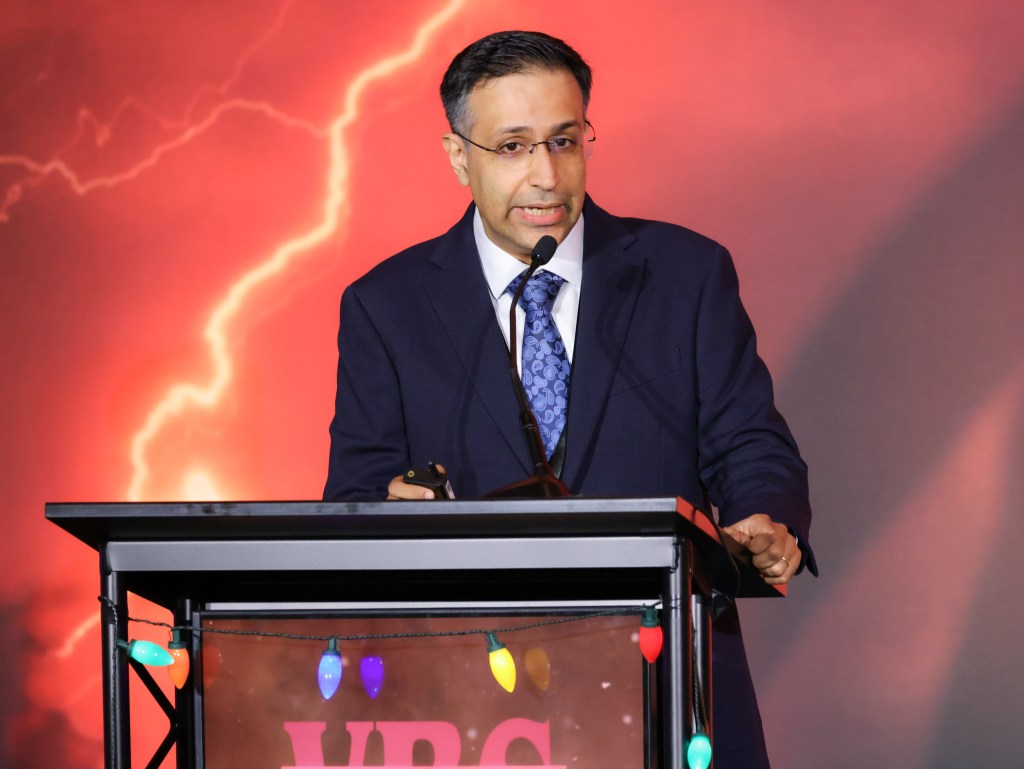
Next, we had the opportunity to learn from Dr. Pradeep Prasad, MD MBA, Associate Professor of Ophthalmology at UCLA Stein Eye Institute and Chief of Ophthalmology at Harbor-UCLA, in his talk entitled Friends Don’t Lie: Tips for Successful TRD Repair. Dr. Prasad emphasizes the importance of understanding of the anatomy preoperatively. Do the Bscan yourself if you can. Then, optimize your view. If you need iris hooks, put them in! If you need to remove the lens, take it out! Stain the vitreous with steroids. Another tip for success is to maintain hemostasis. Dr. Prasad likes to use the backflush instrument to remove hemorrhage or clot. He begins at the edge of the clot where it is thinner and less adherent, and brushes it toward the source of the bleeding, tamponades it, and ultimately removes the clot. He moves onto segmentation, the “core of the surgery”. Dr. Prasad states that we start with separating the midperipheral posterior vitreous from the anterior vitreous. This decreases the risk of creating an anterior break and can help determine whether the hyaloid is elevated. He then reminds us to follow the tissues and to avoid forcing the cutter in. By doing so, we can break large plaques into smaller pieces, and can access the posterior vitreous which helps to lift the hyaloid above and around the plaques. In TRD surgery, Dr. Prasad states that the cutter is the most powerful tool we have. It has many functions including a hook or aspirator. The fluidics of the cutter can be used to our advantage as well. If you want to create turbulence, use a high vacuum setting with low cut rate, whereas if you want to segment in tight spaces, use low vacuum with higher cut rate. Dr. Prasad leaves us with a few quick tips to round out his excellent talk. He highlights the importance of anterior laser, which can often be done best intraoperatively, that dropping the IOP is a nice technique to find bleeders, and that chronic subretinal fluid in TRDs should be followed post-operatively with serial OCT.

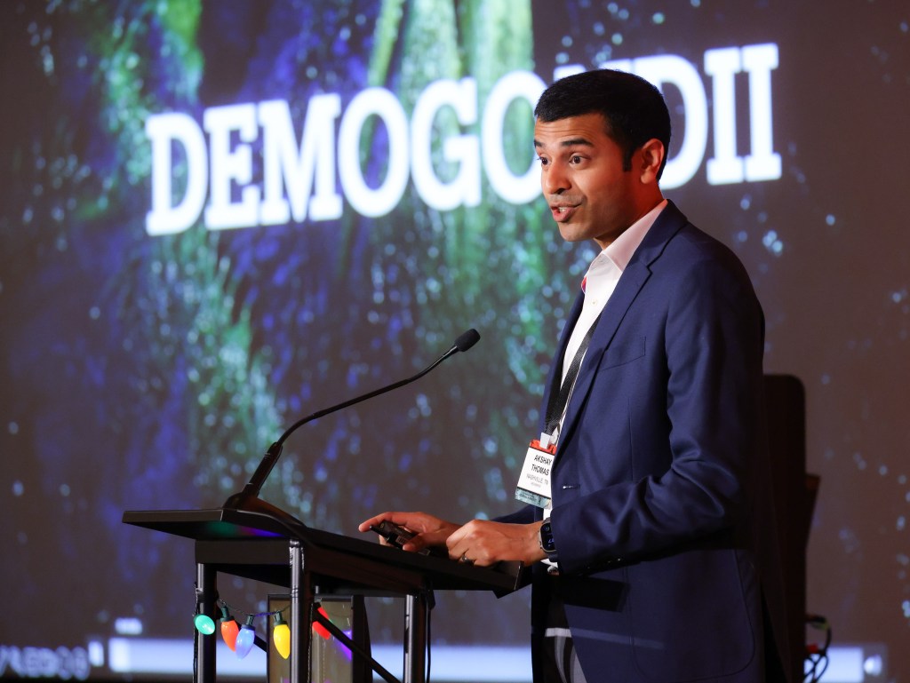
Dr. Akshay Thomas, MD, MS, vitreoretinal surgeon and uveitis specialist at Tennessee Retina, introduces his presentation entitled “Demogondii”, a talk in which he shares surgical and management tips for patients with infectious uveitis. He begins with a case of recalcitrant toxoplasmosis in which laser is applied to the lesion and the area of expansion. This can kill the organism and induce remission. He then describes that in children with infectious macular lesions, there is an increased risk of retinal detachment which can happen after contracture of the regions of scarring. Dr. Thomas states that while it is tempting to peel off the areas of fibrosis intraoperatively, he recommends trimming it instead so as to avoid causing a large retinal hole. His third tip is regarding eyes with fungal infections. He describes that lesions could be preretinal or retinal, and intraoperative OCT can help with this. Dr. Thomas moves on to discuss some tips for retina surgery in patients with infectious keratitis. He recommends careful placement of the surgical wounds, away from areas of prior infection, to suture the wounds to reduce risk of choroidal detachments and hypotony, and consider injection of antivirals after the case to reduce risk of re-activation.

We moved onto a fantastic talk by Dr. Daraius Shroff, MS, FRCS, Medical Director of Shroff Eye Center. Dr. Shroff’s talk is a love letter to the scleral buckle. He starts off by laying some ground rules. We will never regret placing a buckle, but may regret NOT placing one. If there are membranes under the retina, you can still place a buckle. And age is nothing but a number – buckle away! Dr. Shroff then goes on to share a few interesting cases. He begins with a young patient with an inferotemporal macula-on retinal detachment who undergoes non-drainage scleral buckle. One week later, she returned with persistent fluid, with concave borders. In this case, they decided to carefully observe, and, 2 months later, the fluid had resolved without intervention. In a second case, Dr. Thomas describes removing an extruded MIRAgel scleral buckle, a device used in the 1970’s. He details his approach of using simple tools such as a chalazion scoop and a suction cannula to remove the gel-like material. The next interesting case he shares is the use of a scleral buckle in a monocular patient with severe corneal scarring. He describes chandelier illumination to place the buckle, demonstrating that even with a compromised view, a buckle can save the day.

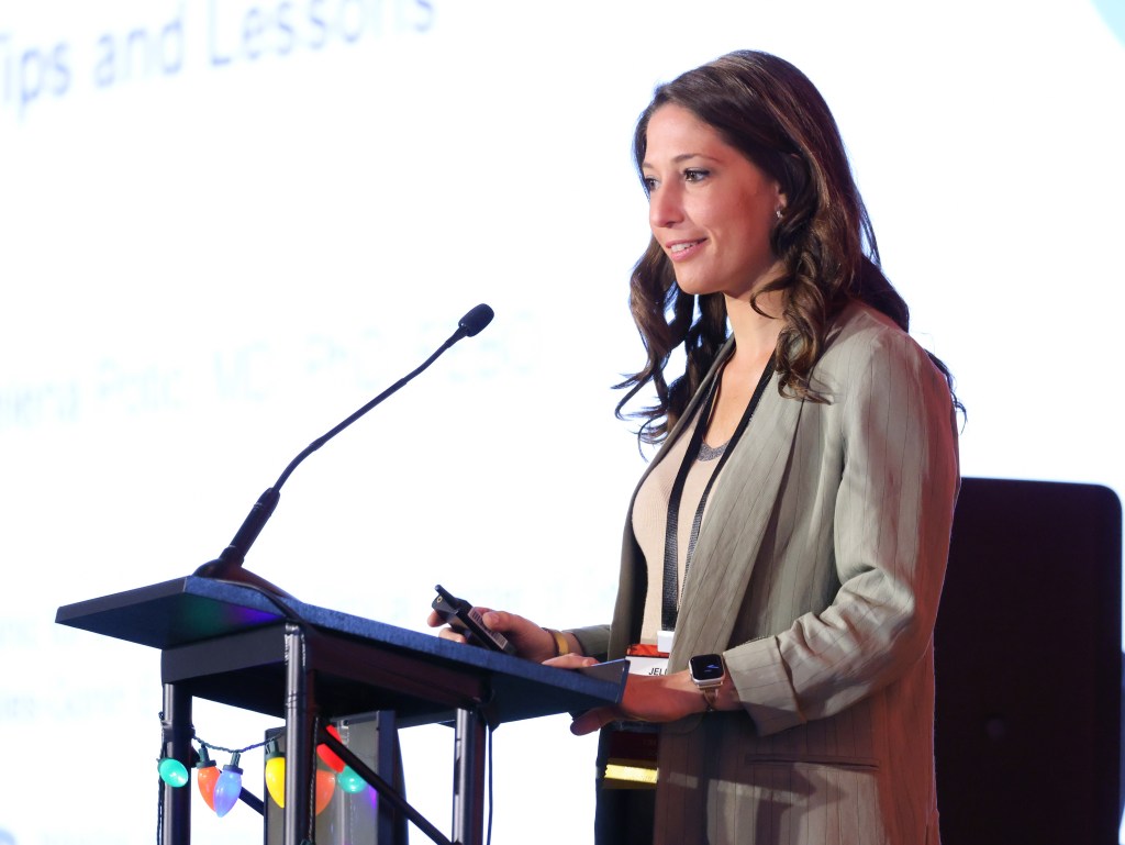
The next talk was entitled “It’s very metal what you did: IOFB in penetrating eye injury – Tips and Lessons” by Dr. Jelena Potic, MD, PhD, FEBO of the Jules-Gonin Eye Hospital of the University of Lausanne, Switzerland. She starts by showing a fascinating surgical video of removing via the retinal break using a metallic intraocular foreign body by using an endomagnet. She emphasizes the importance of the mechanism of injury as well as the time course of the patient’s symptoms, as these factors can have a significant impact on surgical planning. Dr Potic describes a case of intraocular foreign body removal and retinal detachment repair in a young man with light perception vision after trauma. The case presented unique challenges including difficult visualization with vitreous hemorrhage and adherent vitreous in a young person. With careful surgical techniques, however, the patient ultimately attained 20/40 vision, a great reminder to “not give up on these eyes” as stated by Dr. Potic.

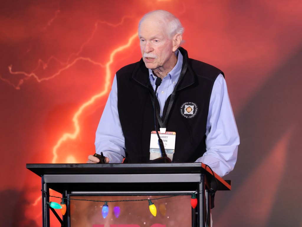
The session came to a close with an interesting and informative talk by legendary Vitreoretinal surgeon, Professor, and J. Donald M. Gass Chair in Ophthalmology at the Bascom Palmer Eye Institute, Dr. Harry W. Flynn Jr., MD. In Dr. Flynn’s talk entitled “Vitrectomy for Retained Lens Fragments After Cataract Surgery: Does Timing Matter?”, he shares the results of their retrospective analysis of patients who had undergone pars plana vitrectomy due to retained lens fragments at differing time points (same day, 1st week, and >1 week after cataract surgery) at BPEI. Interestingly, according to Dr. Flynn’s study, visual acuity and rates of retinal detachment did not differ significantly between the three groups regardless of timing of vitrectomy. In such cases, it may be beneficial to remove the retained lens fragment that day so as to prevent having to place the patient under anesthesia another time. However, this option may not be available to all depending upon the practice setting.

Photographs courtesy of Kevin Caldwell