Chloe Y. Li, MD, MA
Columbia University/New York Presbyterian
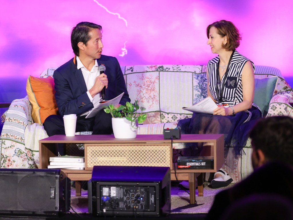
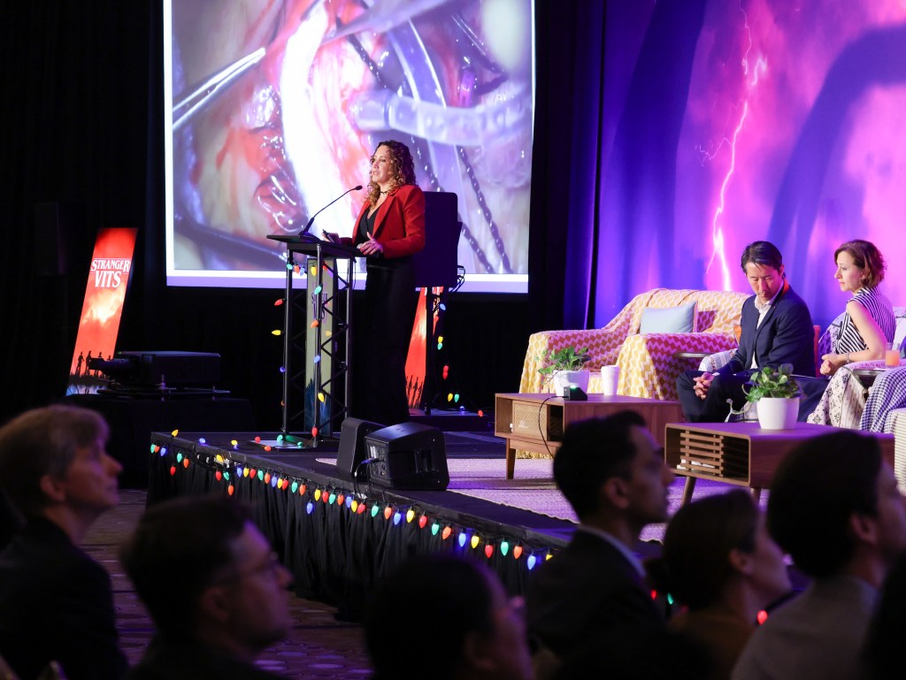
The second-ever “North vs South” debates at Vit Buckle Society featured four retina surgeons from Canada and four from Mexico. Topics included novel management strategies for complications during ILM flap creating, complex retinal detachment repairs, myopic maculopathy surgery, and dissection of proliferative vitreoretinopathy membranes. It was moderated by Dr. Allen Ho, of Wills Eye Hospital, and Dr. Sengul Ozdek, of Gazi University.
Topic 1: Managing ILM Flap Complications
Up first were Drs. Virgilio Morales-Canton, MD (South) and Parnian Arjmand (North) debating on the management of complications during ILM peeling. Dr. Morales-Canton presented a nail-biting case of a patient with a myopic macular hole, for whom he performed an ILM peel with flap. However, during aspiration of residual PFO, the ILM flap was inadvertently also aspirated. The patient then developed a total RRD. During subsequent surgery, Dr. Morales-Canton administered endolaser to the edges of the macular hole, allowing the macular hole to close. The patient’s vision at the final follow up was 20/50. Dr. Ozdek commented it may be possible to use Tisseal glue instead of endolaser in cases of a lost ILM flap.

Dr. Arjmand took a more generalized approach and discussed strategies to avoid four common ILM flap complications: flap amputation, small flap, and aspiration of the flap. She recommended peeling of the ILM flap in a spiral pattern, starting counterclockwise and alternating direction, to create several flaps of varying sizes. This allows the surgeon to sidestep issues of flap amputation, small flap, and flap aspiration by making multiple flaps available of varying sizes. She also advocated for the use of zero suction, use of Tano scraper when faced with difficulty initiating the peel, and use of viscoelastic to fix the flap in place if there is concern for flap movement.
Both speakers emphasized the danger of ILM flap movement or aspiration, and the need for an accessible backup plan once it happens.

Topic 2: New and Old Approaches to Retinal Detachment Repair
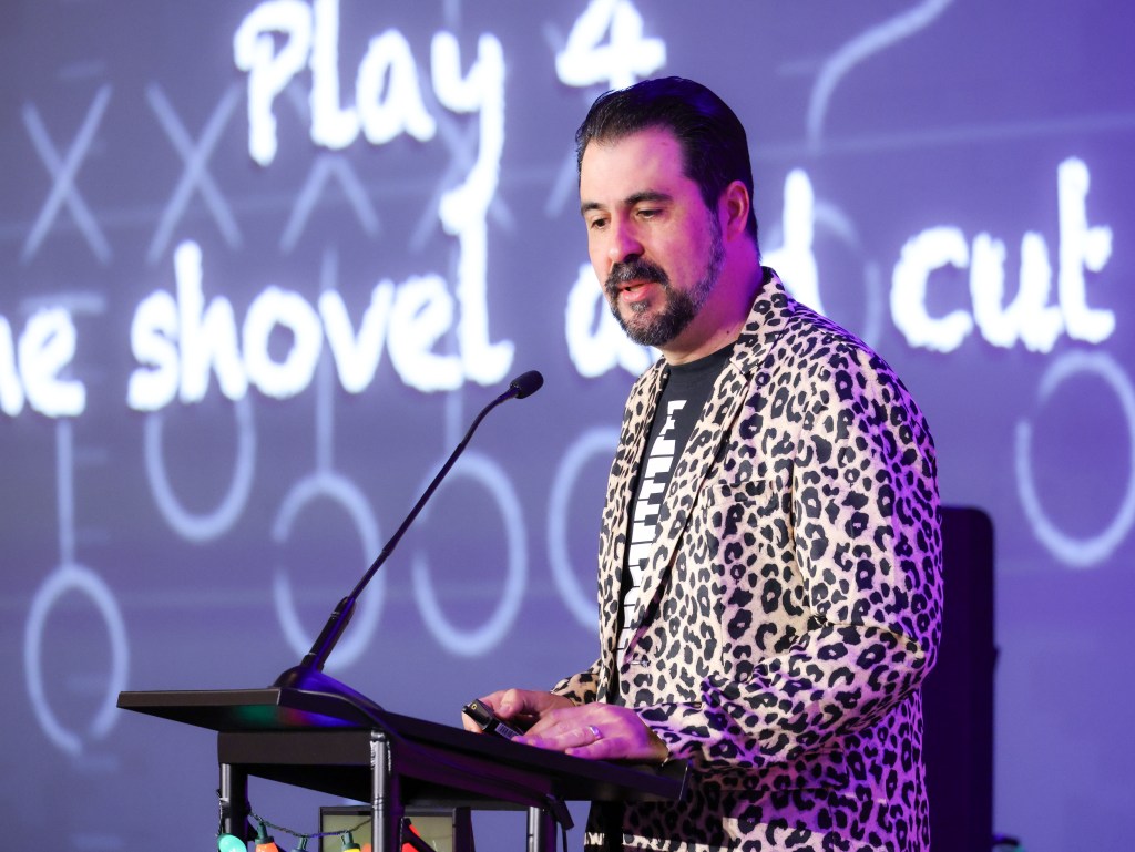
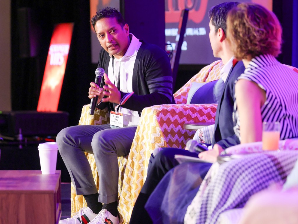
The second set of interlocuters were Drs. Gerardo Garcia-Aguirre (South) and Rajeev Muni (North), discussing two types of challenging retinal detachment repairs. First, Dr. Garcia-Aguirre gave advice on managing advanced diabetic TRDs. To begin, he recommended using as bright a light as possible, followed by complete dissection and removal of tractional membranes, using a “shovel and cut” technique. He reminded us that tractional membranes can extend far into the peripheral retina, and that care must be taken to remove them completely. When cutting through thick membranes, he advocated use of a slow cut rate (1000x). He argued for liberal use of diathermy to minimize new bleeding during surgery, a point strongly seconded by Dr. Ozdek.

Next, Dr. Rajeev Muni described a daring new technique for repair of rhegmatogenous retinal detachments: suprachoroidal viscopexy. He argued that some retina surgeons (American ones, perhaps?) rely too much on scleral buckling and vitrectomy. This is because vitrectomy with gas tamponade and buckling both induce too-rapid retinal reattachment and subretinal fluid resolution, which can lead to retinal folds and displacement. This results in anatomically successful but functionally poor outcomes, as patients experience metamorphopsias and aniseikonia. Instead, he proposed using a 30g needle to inject sodium hyaluronate into the suprachoroidal space adjacent to a retinal break under direct visualization. Sodium hyaluronate is injected until a choroidal indentation is appreciated on indirect ophthalmoscopy. Dr. Muni reported patients had a gradual resorption of SRF with excellent visual recovery after suprachoroidal viscopexy. However, on questioning by Drs. Ho and Ozdek, Dr. Muni conceded he has selected only patients with shallow temporal and superotemporal RRDs for this technique, and that inadvertent retinal puncture has occurred during instillation of the sodium hyaluronate.

Topic 3: Magical Myope Management Strategies
Next, Dr. Gabriela Lopez Carasa (South) and Dr. Flavio Rezende (North) discussed repair of complex RDs in highly myopic eyes. Dr. Lopez proposed using polytetrafluoroethylene (PTFE) felt, a biocompatible, porous polymer used in vascular grafting, as a macular indentation or buckling device. In a patient with myopic tractional maculopathy, she induced hypotony by placing sclerotomy ports, anchored a small piece of PTFE felt with Mersilene suture, and used a Snellen loop to place the felt under the macula. She used intra-operative ultrasound and OCT to place the felt into the precise location of the staphyloma and was able to verify shortening of the patient’s axial length from 36 to 32mm. Following surgery, the patient’s macular SRF was resolved and vision improved.

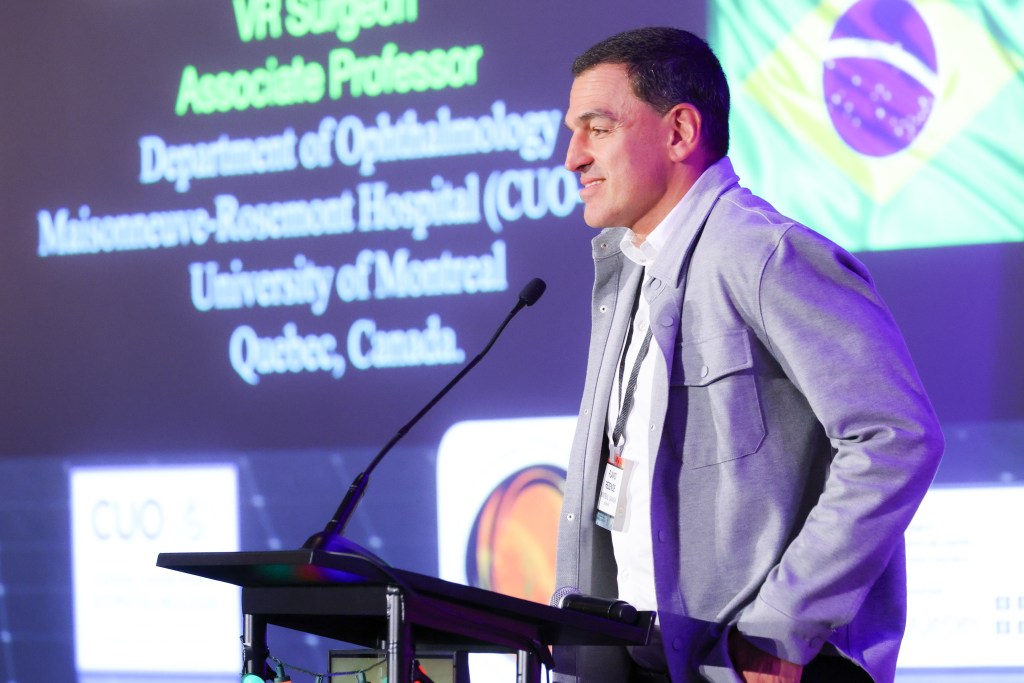
In contrast, Dr. Flavio Rezende argued for use of endoscopy during repair of a re-detachment under silicone oil in a highly myopic and hypotonous eye. First, he performed routine vitrectomy and careful bimanual dissection of thick PVR membranes under oil, then ILM peel. He used endoscopic dissection to remove a fibrous ring of anterior PVR that had formed at the sites of the sclerotomies made during the first detachment repair. The endoscopic view allowed for direct visualization and verification of ciliary body function. He argued that the removal of anterior PVR and verification of ciliary body function would not have been possible with standard vitrectomy. Post-operatively, the patient underwent a series of weekly methotrexate injections; he had a final VA of 20/300 with normal IOP under silicone oil.
In highly myopic eyes prone to complication, both speakers had unique solutions.

Topic 4: Very Vascular Considerations
Finally, Dr. Maria A. Martinez-Castellanos (South) and Dr. Efrem Mandelcorn (North) gave pearls on the management of vascular retinal diseases in children and adults. Dr. Martinez-Castellanos, a pediatric specialist, highlighted that surgical management of diseases of vascular development, such as ROP or FEVR, the anterior hyaloid must be completely removed. In cases when the hyaloid cannot be separated from the lens or iris, these structures should be sacrificed to prevent development of anterior proliferative disease in children. Her takeaway: once anterior proliferation has begun in a pediatric patient, it is highly contractile and aggressive, and inevitably leads to detachment.

Dr. Mandelcorn described a different vascular issue in adults; he used the neovascular proliferative tissue growing from the retina of a diabetic patient to plug a macular hole without any gas or oil tamponade. The patient remained attached briefly, but then developed recurrent hole. He then used an amniotic membrane graft approach. With the cutter on reflux mode, he created a “shelf” between the retina and RPE at the edges of the macular hole and then tucked an amniotic membrane graft into the shelf with the stromal side down. Again, this was temporarily successful, but the patient eventually developed another macular hole and detachment. Finally, Dr. Mandelcorn used two retinal autografts, harvested from the patient’s nasal retina, to close this macular hole. After 4 macular grafts, the patient’s macular hole finally remained closed with 20/200 vision.
During this last discussion, the North and South both showed us that (neo)vascular tissue during retina surgery is a tricky beast indeed.

Conclusion:
The audience voted by QR code and found that the North won by a vote of 65% to 35%. However, the most valuable lesson of the day came from Dr. Garcia Aguirre, who proclaimed: “Always avoid fellow-induced foveophagia during retinal surgery!”
Photos courtesy of Kevin Caldwell