Kotaro Tsuboi, MD
Aichi Medical University, Nagakute, Japan
The second day of Fuji Retina 2024 featured a captivating afternoon dedicated to scientific discussions on vitreoretinal imaging and vascular disease. Below is a summary of selected papers from the session.
Innovations in Choriocapillaris Imaging:
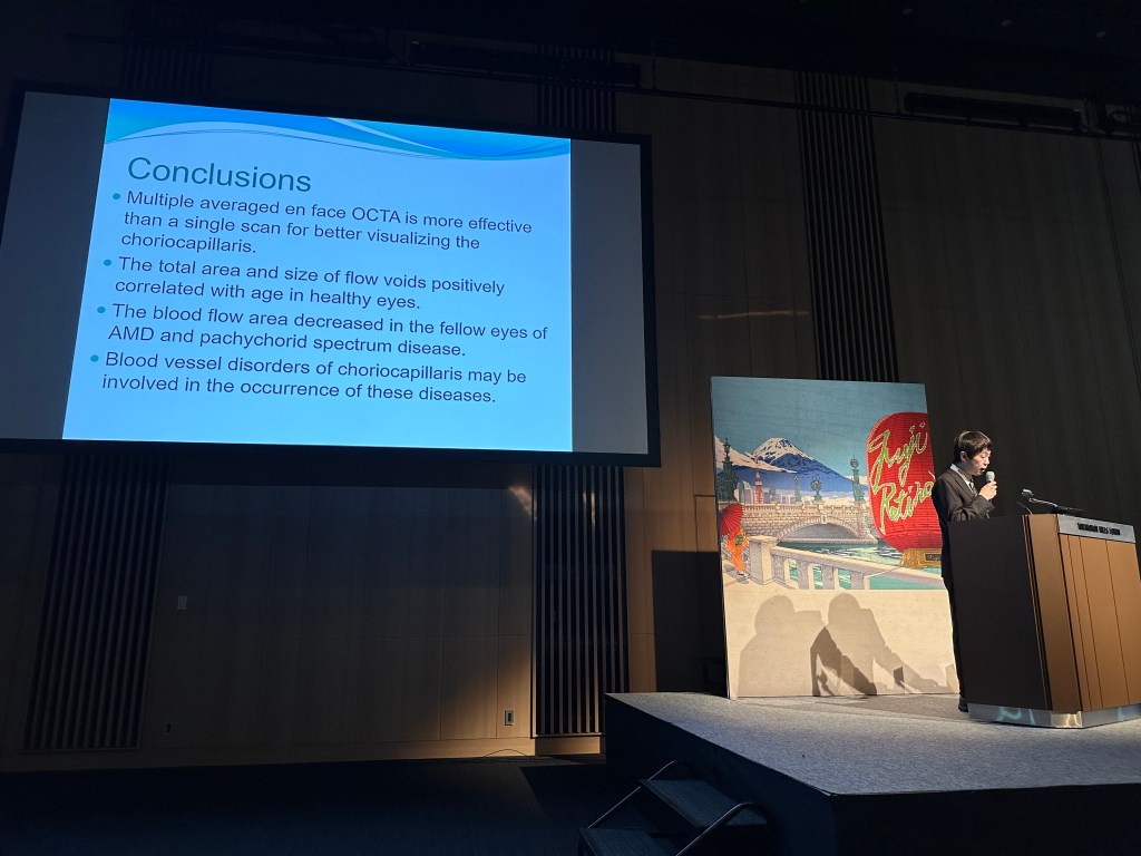
Dr. Sotaro Ooto from Kyoto University, Japan, kicked off the session with his presentation titled “Choriocapillaris Imaging by Averaging Multiple En Face OCTA Images.” Dr. Ooto introduced a novel technique for acquiring high-resolution images of the choriocapillaris by averaging out data from nine individual 3×3-mm OCTA scans. This method allowed for a detailed analysis of the choriocapillaris, showing a correlation between the area and size of flow voids with aging in healthy individuals. Furthermore, a decrease in blood flow area was observed in individuals with one eye affected by age-related macular degeneration (AMD) or pachychoroid spectrum disease, suggesting an early involvement of choriocapillaris blood vessel disorders in these diseases. Dr. Ooto’s findings highlight the importance of using multiple averaged en face OCTA scans for a clearer visualization of the choriocapillaris.
Ultra-Widefield Imaging’s Role in Retinal Vascular Diseases:
Next, Dr. Adrienne Scott from the Wilmer Eye Institute at Johns Hopkins University School of Medicine, USA, presented insights into the utility of ultra-widefield imaging in retinal diseases and provided an update on the management of proliferative sickle cell retinopathy. Dr. Scott underscored the advantages of ultra-widefield (UWF)1 imaging over traditional methods, particularly for diagnosing and managing retinal vascular diseases. Dr. Scott explained that UWF imaging provides a broader view of the retina, uncovering important details about conditions such as diabetic retinopathy, which are often overlooked by standard imaging techniques. She referred to the DRCR Protocol AA findings, which demonstrated that predominantly peripheral lesions (PPL) identified through UWF color retinal imaging did not predict a significant, 2-step deterioration on the Diabetic Retinopathy Severity Scale (DRSS).2 Dr. Scott’s discussions revealed how UWF imaging offers a comprehensive view of the retina, aiding in the detection and treatment guidance of conditions such as diabetic retinopathy. Despite its benefits, challenges like image distortion, the need for specialized knowledge, and higher costs were acknowledged. She also explored the potential future applications of UWF imaging in improving our understanding of retinal diseases and its integration into artificial intelligence and screening programs.


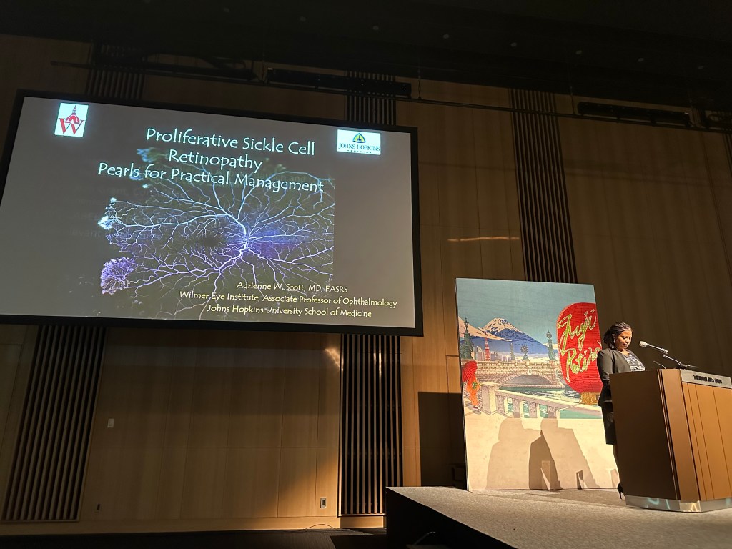
Advances in Sickle Cell Retinopathy Management:
In her second talk, Dr. Scott focused on advancements in sickle cell retinopathy management, highlighting the classification system introduced by Dr. Goldberg in 1971 that remains in use today. This system identifies stages of vascular remodeling in the retinal periphery, notably starting with peripheral arterial occlusions and progressing through stages that include the development of sea fan-shaped vascular formations. Sickle cell retinopathy, which varies in severity among different genotypes of the disease, shows a higher prevalence in the hemoglobin SC genotype. This condition primarily affects adolescents and young adults and is characterized by a unique occurrence where up to 40% of sea fan neovascular formations may spontaneously regress without leading to vision loss. The presentation also covered the challenges in screening and treatment for sickle cell retinopathy, including the timing and choice of therapeutic interventions like laser therapy and anti-VEGF treatments. Despite a lack of consensus on treatment strategies, laser photocoagulation remains a primary option, significantly reducing the risk of vitreous hemorrhage without preventing new sea fan formation. Dr. Scott highlighted case studies indicating that VEGF is released not just from areas of non-perfusion but also from zones transitioning between perfused and non-perfused regions. This research suggested that laser treatments should target these transitional zones where VEGF concentrations are highest. Dr. Scott concluded by emphasizing the importance of personalized treatment approaches, including the use of anti-VEGF therapy and surgery in advanced cases, to improve outcomes for patients with sickle cell retinopathy. Her presentation emphasized the intricate nature of treating sickle cell retinopathy and the ongoing need for research to refine therapeutic strategies.

Distinguishing Placoid Disorders through Imaging
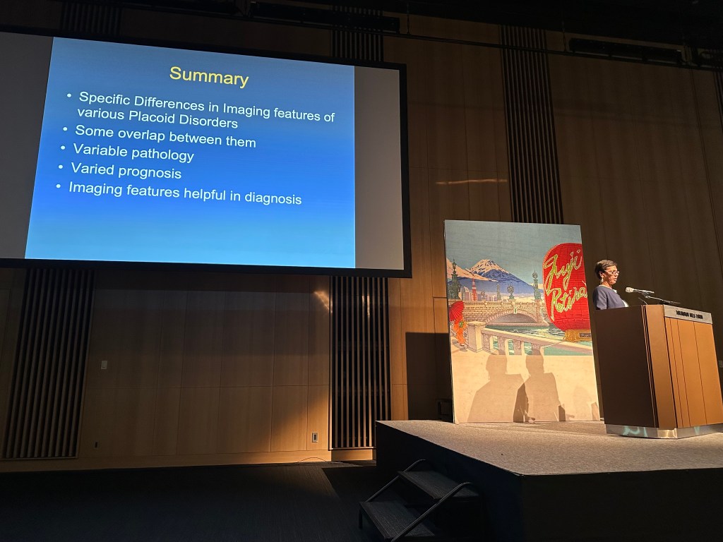
Dr. Anita Agarwal from West Coast Retina & Vanderbilt Eye Institute, USA, offered an in-depth look at “Imaging Differences of Placoid Disorders,” emphasizing the essential role of multifocal imaging techniques in diagnosing conditions such as acute posterior multifocal placoid pigment epitheliopathy (APMPPE), tuberculosis (TB)-related serpiginous choroiditis, placoid syphilis, and persistent placoid maculopathy. She highlighted how advancements in imaging technologies, including fluorescein angiography, OCT and OCT angiography, have become invaluable in detecting and understanding diseases that were previously challenging to diagnose and manage.

Multimodal Imaging in Macular Telangiectasia Type 2:
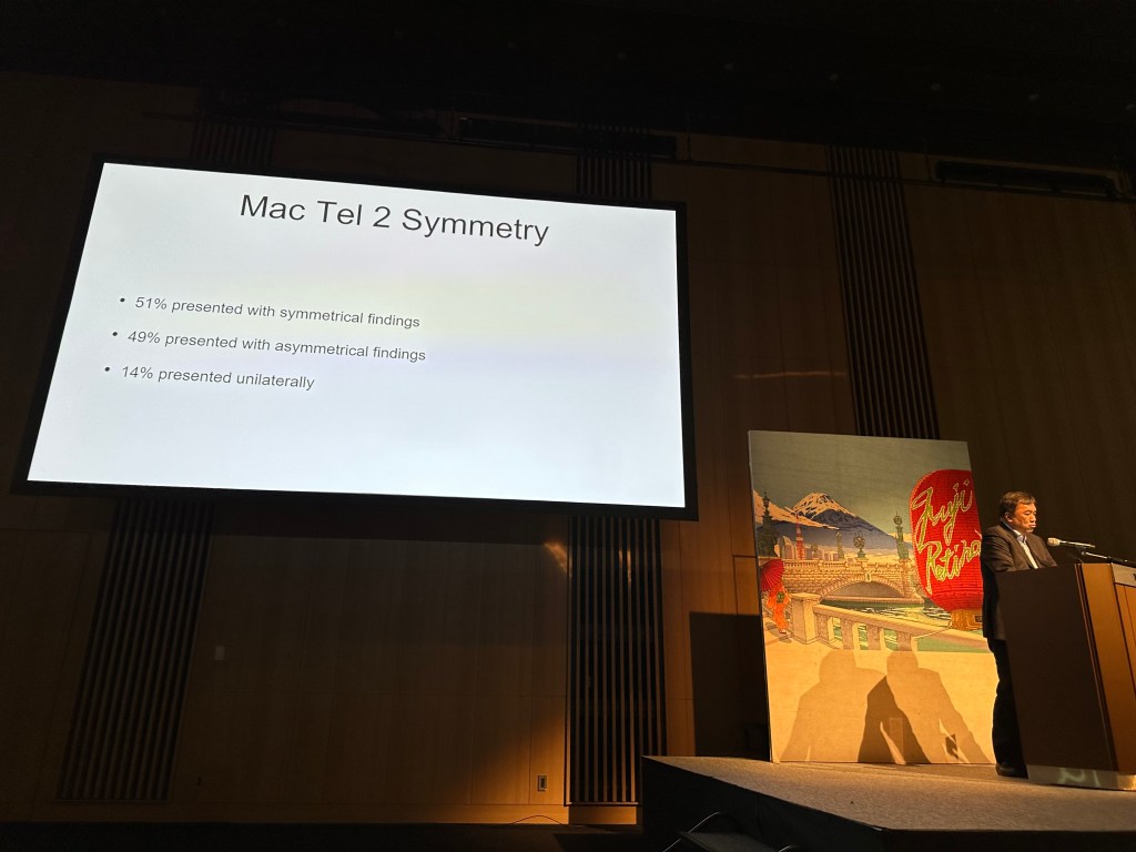
Dr. Lihteh Wu from Asociados de Macula, Vitreo y Retina de Costa Rica presented his findings on “Multimodal imaging in macular telangiectasia 2,” focusing on a cohort study from Latin America. The study encompassed 182 patients, predominantly female (mean age 62, range 25 to 80), shedding light on the varied presentations and multimodal imaging findings associated with MacTel type 2. The patients in the study displayed a wide range of visual acuities and findings from multimodal imaging, such as changes in pigment, fluorescence, hyperreflectivity, shadows on the retina, and flow mapping seen through various imaging techniques. Particular focus was placed on the absence of normal luteal hyporeflectance and the detection of features like cavitations in the inner and outer layers of the retina, crystals, expanded blood vessels, and increased fluorescence. Among the most significant clinical observations were the expanded retinal blood vessels, with abnormalities in fundus autofluorescence being one of the earliest signs identified. The research illustrated how the disease’s stage, based on the classification by Chew et al.,3 correlates with visual acuity. Additionally, the study investigated whether the disease manifested symmetrically in both eyes of the patients, discovering that 51% of the cases showed no difference in disease stage between the two eyes, indicating symmetry. The remaining cases either exhibited asymmetrical stages of the disease across the two eyes or showed no signs of the disease.

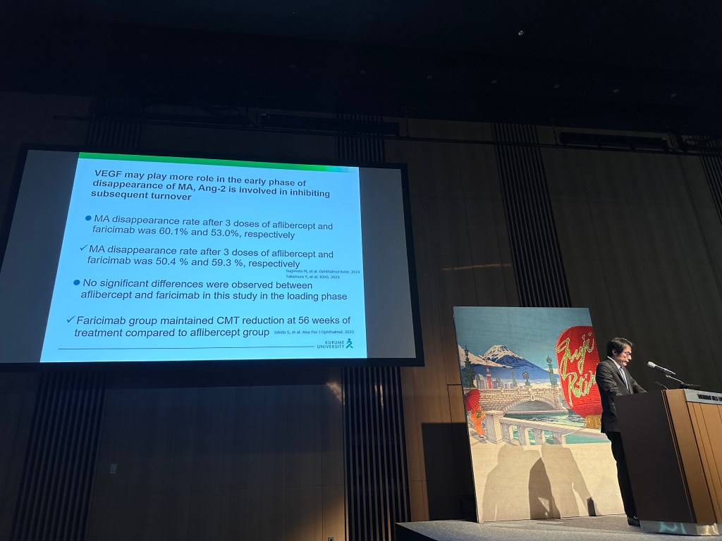
Comparing Treatments for Microaneurysms in Diabetic Macular Edema:
Dr. Shigeo Yoshida from Kurume University, Japan, presented “Microaneurysms in the Treatment of DME.” He reported on a study evaluating the effect of intravitreal faricimab (IVF) on the reduction of microaneurysms (MA) compared with those treated with intravitreal aflibercept (IVA). Both the IVA and IVF groups demonstrated a significant reduction in central macular thickness (CMT) and improvement in visual acuity after three injections. Regarding the number of MAs, over 50% disappeared after three consecutive injections, yet no differences were observed in CMT, visual improvements, or the reduction of MAs between the two groups.

This session underscored the dynamic advancements in retinal imaging and the management of vascular diseases, offering new insights and reinforcing the importance of continual research and innovation in the field.
References
- Choudhry N, Duker JS, Freund KB, et al. Classification and Guidelines for Widefield Imaging: Recommendations from the International Widefield Imaging Study Group. Ophthalmol Retina. 2019;3(10):843-849. doi:10.1016/j.oret.2019.05.007)
- Marcus DM, Silva PS, Liu D, et al. Association of Predominantly Peripheral Lesions on Ultra-Widefield Imaging and the Risk of Diabetic Retinopathy Worsening Over Time [published correction appears in JAMA Ophthalmol. 2023 Jan 1;141(1):104].JAMA Ophthalmol. 2022;140(10):946-954. doi:10.1001/jamaophthalmol.2022.3131
- Chew EY, Peto T, Clemons TE, et al. Macular Telangiectasia Type 2: A Classification System Using MultiModal Imaging MacTel Project Report Number 10. Ophthalmol Sci. 2022;3(2):100261. Published 2022 Dec 8. doi:10.1016/j.xops.2022.100261