Melissa Yuan, MD
Ophthalmology Resident
Massachusetts Eye and Ear Infirmary
The 2024 conference finished strong with the medical and surgical case conferences.
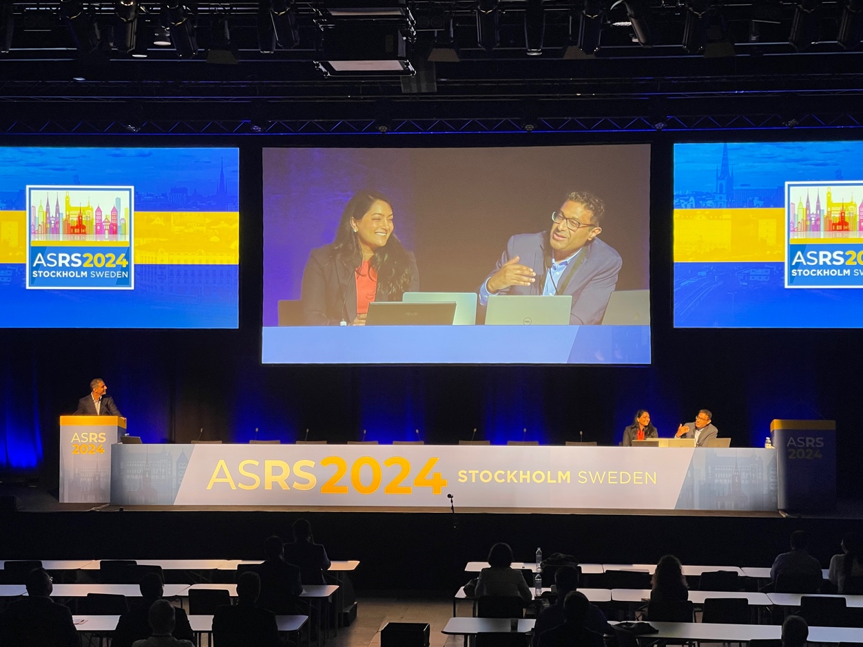
The medical case conference session was moderated by Drs. Sruthi Arepalli and Srinivas Sadda and featured cases by Drs. J Fernando Arevalo, Narciso Atienza, Sreenivasa Basavanthappa, Paul Bernstein, Rachel Downes, Jaclyn Kovach, Yasha Modi, Mohamed Mohamed, Kareem Moussa, Kshitij Raizada, Avik Sarkar, Mohammad Siddiqui, Bradley Smith, and Scott Walter.
Dr. J. Fernando Arevalo kicked off the session with a case entitled “Yellow Spots,” of a healthy elderly patient with no past ocular history or symptoms, found to have multiple yellow creamy lesions and choroidal thickening. The diagnosis was ultimately determined to be benign reactive lymphoid hyperplasia after negative workup including vitreous biopsy and PET scan. Dr. Sadda brought up the work of Drs. Jasmine Francis and David Abramson, which postulates an antigenic stimulus resulting in this immune reaction, and inquired about steroid or antibiotic treatment. This patient had declined all treatment due to his lack of symptoms.
 Dr. Narciso Atienza then showed a number of stunning photographs in his talk “A Mystery Tumor,” featuring a young patient with a large solid mass compressing the globe which was determined to be a tuberculoma on histopathology. He interestingly had negative systemic tuberculosis testing via QuantiFERON Gold testing. Subsequently, Dr. Sreenivasa Basavanthappa’s case “Unmasking the Great Masquerader” followed a patient referred for post-cataract surgery macular edema which responded well to intravitreal dexamethasone. Unfortunately, the intravitreal steroids precipitated acute vision loss in the left eye, with a large placoid chorioretinal lesion, focal thickening and nodularity of the RPE with disruption of the overlying ellipsoid zone, confirmed to be syphilitic chorioretinitis. The patient was treated with IV penicillin with good response. Dr. Arepalli raised two important teaching points: ruling out infection before steroid administration, and that the outer retinal changes and nodularity in ocular syphilis are a good way to follow response to treatment as they typically improve quickly.
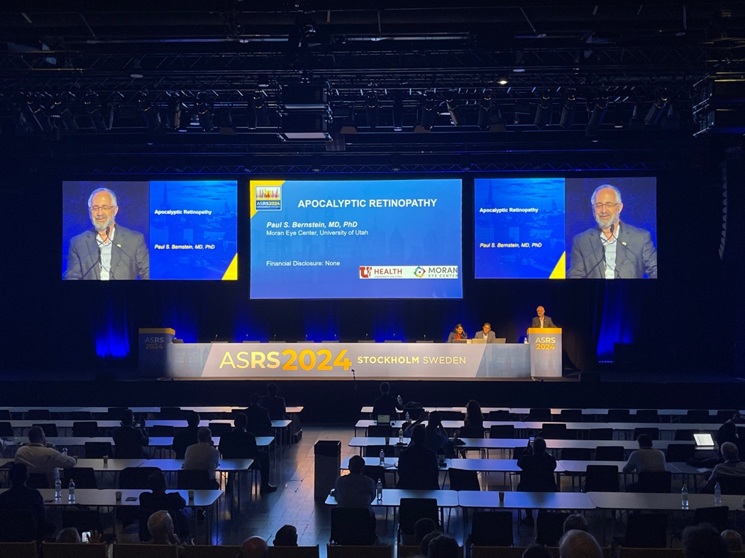
Then, Dr. Paul Bernstein presented a startling case entitled “Apocalyptic Retinopathy,” of a young woman with loss of vision in both eyes in the setting of a suicide attempt with a large number of anti-radiation tablets. Visual acuities were significantly reduced with unrecordable ERGs and complete wipeout of outer retina and RPE. The diagnosis was potassium iodate retinopathy. He taught the audience that potassium iodate is a well-known RPE toxin used in animal models of retinal degeneration, but also an approved agent in the US to iodize salt (at much lower doses) and marketed as antiradiation tablets to be consumed in the case of a nuclear disaster. Importantly, this compound has a very narrow therapeutic window. An audience member commented on two prior cases in the literature of patients receiving unintentional systemic administration of sodium iodate, causing severe macular toxicity – not the entire retina as in Dr. Bernstein’s case – highlighting the sensitivity of the macula to these oxidizing agents.

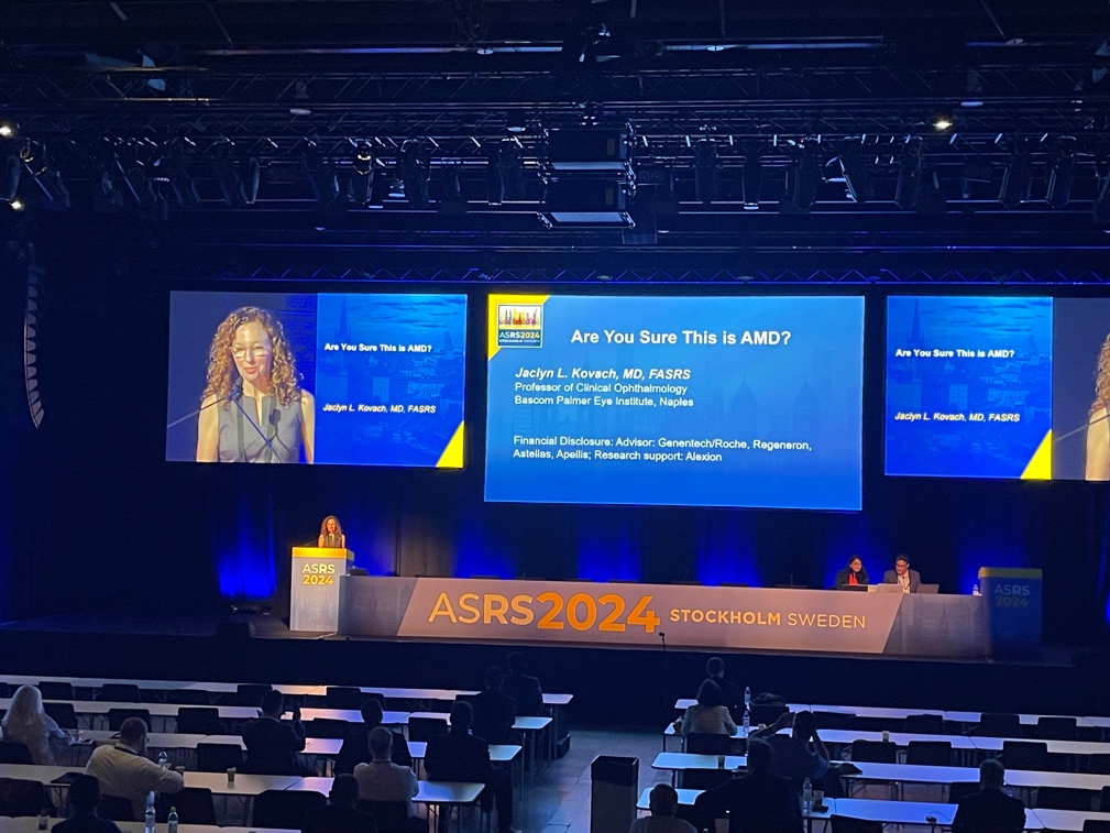
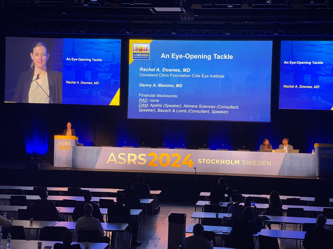
Dr. Rachel Downes presented a case of whiplash maculopathy after a football tackle with prominent bilateral bacillary layer detachments that improved spontaneously, aptly entitled “An Eye-Opening Tackle.” This was followed by Dr. Jaclyn Kovach’s case, “Are you sure this is AMD?”, of a middle-aged man with a presumed diagnosis of AMD with subretinal fibrosis in one eye and reticular pseudodrusen and subretinal deposits in both eyes, later diagnosed with Sorsby Fundus Dystrophy on genetic testing. Dr. Sadda asked whether Dr. Kovach would obtain genetic testing on patients with extensive peripheral drusen. Dr. Kovach answered that she does not routinely, but this case’s advanced presentation in a relatively young patient led her to seek alternate diagnoses.
 Dr. Yasha Modi shared “An Immunologic Paradox”, a case of a young female with common variable immunodeficiency who was diagnosed with severe VKH. The audience was reminded that CVID can have paradoxical autoimmunity in up to half of patients due to T cell dysregulation, despite it being an immunodeficient state. Switching gears, Dr. Mohamed R Mohamed presented a case of orbital compartment syndrome after scleral buckle. While IOP elevation has been seen after scleral buckling due to angle closure, this case was postulated to have a different mechanism, possibly from retrobulbar hemorrhage due to the peritomy or injury to the vortex veins. Dr. Kareem Moussa followed with a case of presumed VKH in a young man with a recent viral infection presenting with decreased vision, erythematous discs, and pockets of subretinal fluid. The patient, however, was finally diagnosed with central serous chorioretinopathy (likely precipitated by steroid inhalers for the URI) based on history and multimodal imaging, as well as hypervitaminosis A leading to intracranial hypertension. This was an excellent example of Hickam’s Dictum. A point that was raised by moderator Dr. Sadda in several cases, including the cases by Drs. Downes, Modi, and Moussa, was the importance of inspection of the choroid. Specifically, Dr. Sadda mentioned that the vessel profiles were not well-visualized on OCT in cases of infiltrative etiologies such as VKH, but they were well-
  defined without opacification in Dr. Moussa’s case of CSR.
Dr. Kshitij Raizada presented a case of a large retinal hole following laser injury, with impressive photos of a large retinal hole oozing fresh blood in the vitreous due to inadvertent Nd:YAG laser damage to the peripheral retina (the laser had been intended for cosmetic purposes). Then, Dr. Avik Sarkar showed a case of electric shock retinopathy that presented asymmetrically one month after electrocution. Changing the topic from lasers and electricity, Dr. Mohammad Siddiqui shared a case of Wolfram-like syndrome, a less-common and more recently described autosomal dominant condition characterized by optic atrophy and deafness and less commonly associated with the diabetes insipidus/mellitus seen in Wolfram syndrome. The laminated OPL as seen in this case was a clue to the diagnosis.
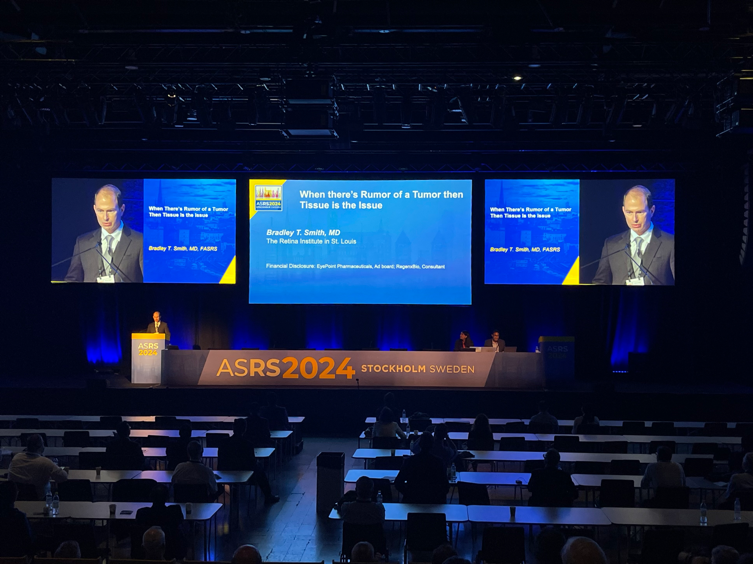

A poetically entitled case by Dr. Bradley Smith, “When there’s rumor of a tumor then tissue is the issue,” took the audience through the case of a ring-like mass lesion infiltrating the choroid, ultimately taken to biopsy by Dr. Smith and luckily found to have extrascleral extension. Pathology confirmed the diagnosis of a low-grade B-cell non-Hodgkin lymphoma. Continuing on the theme of neoplasms, Dr. Scott Walter presented a case of “Neoplastic Proliferative Vitreoretinopathy” in a patient with vitreoretinal metastases of cutaneous melanoma, with clinical course complicated by a PVR detachment and subsequent methotrexate resistance requiring intravitreal topotecan. Dr. Arepalli asked about the decision-making regarding the choice of topotecan, and Dr. Walter explained that it is more stable than intravitreal melphalan, which had been his first choice but determined to be impractical for the office setting. Dr. Sadda commented that these types of metastatic and resistant cases are becoming increasingly common in the era of checkpoint inhibitors, possibly due to the immune privilege of the eye making immune checkpoint inhibitors less efficacious in the intravitreal and retinal compartments.
The day than transitioned to the surgical case conference.
The surgical case conference moderators were Drs. Maria Berrocal and Gaurav Shah, and the case presenters included Drs. Nishikant Borse, Jinghua Chen, Srinivas Joshi, Eric Hung, Jayant Kumar, Ananth Sastry, Vinay Shah, Veer Singh, and Jennifer Tingley.

Dr. Nishikant J Borse started the session off with a surgical video of dislodging an embolus using low IOP vitrectomy and arterial massage in CRAO. ILM peeling from macula to disc/peripapillary area was performed, the IOP was lowered, and disc pulsations were seen as the embolus moved toward the disc. The disc was then massaged directly with forceps or soft tip, and pieces of the embolus were visualized moving down the arterioles into the periphery. The possibility of using intraoperative fluorescein angiography was raised by Dr. Berrocal, as this could help assess reperfusion.

Dr. Jinghua Chen showed the removal of a human amnion/chorion membrane graft after closure of a macular hole. This was a case of a patient who had underwent macular hole repair via 25g
PPV with dehydrated human amnion/chorion and silicone oil. 6 months later, as the patient was bothered by the large scotoma due to the membrane, he underwent silicone oil and membrane removal. A Flex Loop was used to lift edge of the membrane and MaxGrip forceps to lift the membrane.
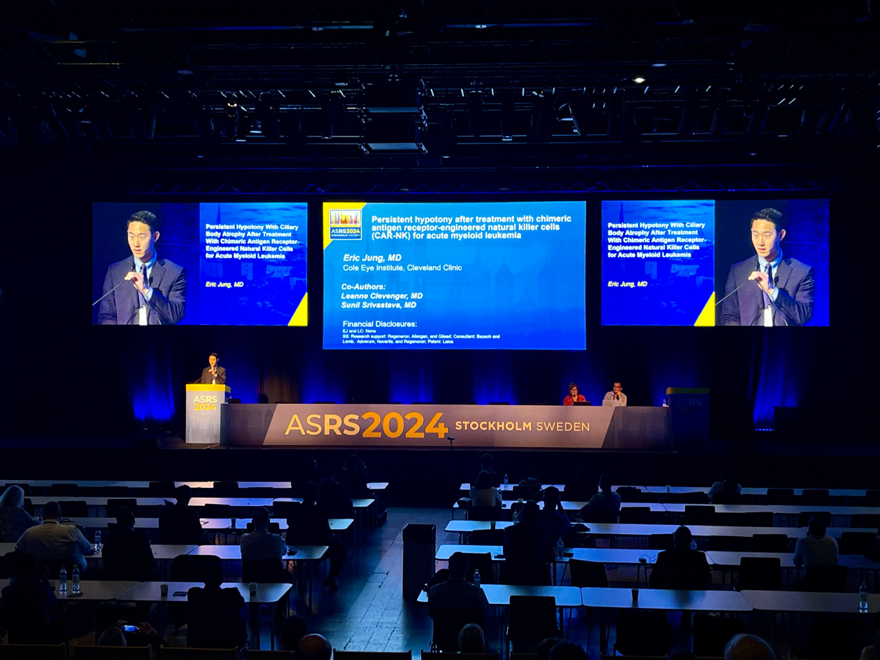

Dr. Srinivas Joshi then presented a case of recreational laser injury with sub-ILM hemorrhage treated with vitrectomy, PVD induction and ILM peeling. Dr. Joshi utilized the monochrome filter for visualization even with less staining of the ILM. The sub-ILM hemorrhage was dislodged gently after ILM peel using a back flush technique. Dr. Shah brought up that this was a helpful technique for macroaneurysms as well. This was followed by Dr. Eric Jung’s case, which combined medical and surgical elements, in patient with persistent hypotony due to ciliary body atrophy after treatment with chimeric antigen receptor engineered natural killer cells for acute myeloid leukemia. The surgical video demonstrated the Retisert surgical implantation and showed the obliteration of the ciliary processes. Both Drs. Shah and Berrocal highlighted some of the difficulties with implantation of the Retisert, with experience gained in the past from the ganciclovir implant, including ensuring the implant is not put in the suprachoroidal space and to close the sclera very meticulously to prevent the possibility of extrusion.

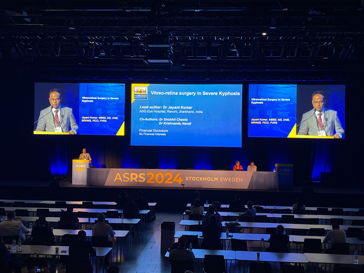
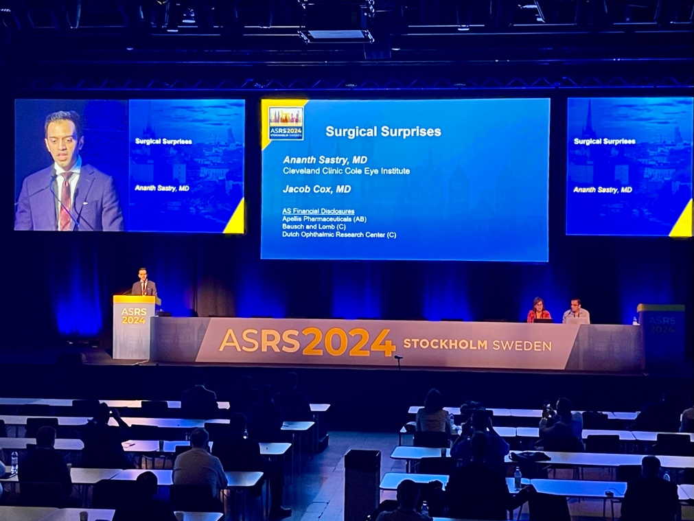
Dr. Jayant Kumar presented on vitreoretinal surgery in severe kyphosis, with attention to the various challenges in positioning and surgeon comfort. Dr. Berrocal commented on her appreciation for these tips as a short surgeon, highlighting the temporal approach and using 3D viewing systems. Then, Dr. Ananth Sastry showed a case of “surgical surprises” in a second opinion recurrent RD repair with a dense cataract and no prior notes. This case features a series of surprises including a residual PFO bubble and gas bubble, a dense posterior hyaloid that required peeling, and a macular hole. There was spirited discussion on how to approach difficult cases such as these, including whether to attempt phacoemulsification first or pars plana lensectomy as in this case; how far out to peel the ILM in these cases; and whether or not to let the PVR membranes mature.

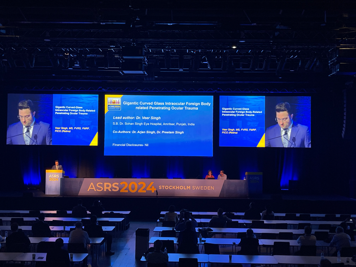
Subsequently, Dr. Vinay Shah demonstrated how four-point bimanual 25-gauge vitrectomy can enhance teaching and fellow performance in complex cases with two surgical videos, one with a more novice fellow and one more experienced. The audience and moderators commented on other teaching modalities including 3D heads up displays. The session and conference ended with two excellent trauma cases. Dr. Veer Singh showed the removal of a very large glass intraocular foreign body, and Dr. Jennifer Tingley shared a video featuring removal of an intraocular BB pellet fragment by standard 3-port vitrectomy.