Elham Sadeghi, MD1, Bita Momenaei, MD2, Justin Muste, MD3, Timothy Xu, MD4
1. Department of Ophthalmology, University of Pittsburgh, School of Medicine
2. The Retina Service of Wills Eye Hospital, Wills Eye Physicians-Mid Atlantic Retina, Thomas Jefferson University, Philadelphia, PA
3. Cleveland Clinic Cole Eye Institute, Cleveland, OH
4. Mayo Clinic, Rochester, MN
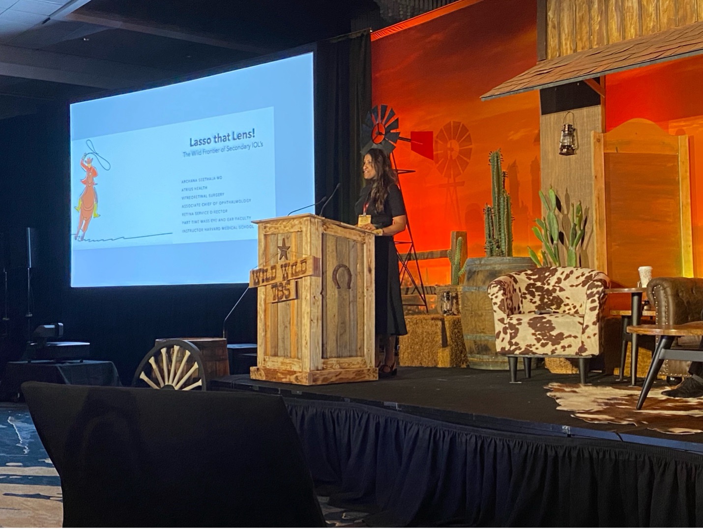
Lasso that Lens – Archana Seethala, MD
Dr. Archana Seetha, MD, reviewed secondary IOLs including the Akreos lens and the cannula-based Yamane technique. Various lenses – MA60, CT Lucia and AR40 – used in the Yamane technique were discussed. While no lens is perfect, selecting the right one and setting patient expectations are crucial. Key steps to implantation were reviewed and troubleshooting was discussed including handing of suture tangling for the Akreos and haptic breakage and unstable haptic-optic junction for the Yamane.
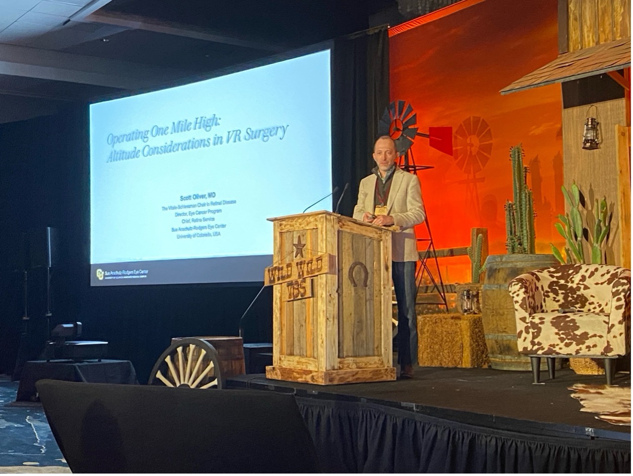
Operating One Mile High: Altitude Considerations in VR Surgery – Scott Oliver, MD
Dr. Scott Oliver, MD, reviewed challenges of retinal surgery at high altitudes. Venturi-based instruments perform less efficiently at altitude, and gas-filled eyes can experience dangerous pressure spikes. Studies show that IOP increases by approximately 11 mmHg per 1,000 feet of ascent, making careful travel planning essential. If patients must fly, then surgeons may place silicone oil instead of gas. Otherwise, patients may fly once the gas bubble decreases to 5% of posterior chamber volume. Strategies to mitigate altitude related IOP increases include prescribing acetazolamide or IOP-lowering drops, mapping lower-altitude driving routes, and periodic rest-stops for every 1,000 feet in elevation to allow for IOP equilibration.

Approach to Biopsies (Vitreous, Choroidal, Retinal Biopsies) – Phoebe Lin, MD, PhD
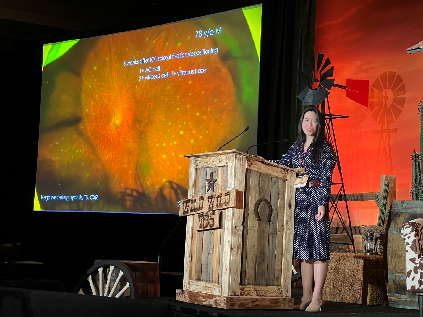

Dr. Lin presented two cases that highlighted the importance of proper surgical technique when pursuing diagnostic vitrectomy and of interdisciplinary collaboration in managing complex vitreoretinal conditions. She showed a case of intraocular lymphoma in a 78-year-old man that required aspiration of stuck-on epiretinal deposits and a case of sarcoidosis in a 78-year-old woman that required chorioretinal biopsy for diagnosis. Although invasive, these biopsies may be necessary in select cases to provide definitive tissue diagnosis when workup is inconclusive. Dr. Lin’s surgical pearls included the importance of preoperative planning such as specimen planning (e.g., coordination with cytopathology and hematopathology, avoiding corticosteroid exposure to maximize diagnostic yield), intraoperative techniques (e.g., Luer-lock syringes to prevent loss of small-volume specimens) and postoperative considerations (e.g, interpretation of pathology results as false negatives are common especially in lymphoma cases where steroids may confound findings).
A Buckle Ain’t Just for Cowboys! How to Start if You’re a Tenderfoot – Chirag D. Jhaveri, MD
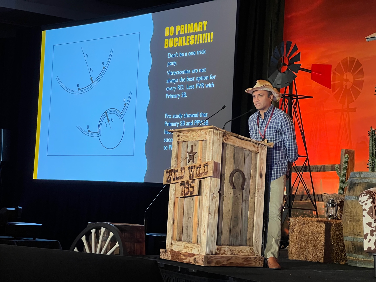

Dr. Jhaveri highlighted the importance of scleral buckling in various clinical scenarios and discussed how scleral buckling is a “dying art” that we must continue practicing, teaching, and learning. The first case was a 67-year-old pseudophakic patient with multifocal IOLs and superior/inferior tears. Given the patient’s high refractive expectations, a radial element buckle was performed to preserve his desired refraction. The second case was a 24-year-old phakic patient with chronic inferior detachment treated with a segmental buckle. Dr. Jhaveri discussed how scleral buckling changes force vectors in a unique way, leading the retina to push outward unlike vitrectomy or gas tamponade. For those in training or uncomfortable with buckling, Dr. Jhaveri suggested starting with vit-buckling but treating the buckle portion as if it were a primary buckle. Dr. Jhaveri’s other surgical pearls included choosing easier cases such as single-quadrant breaks, avoiding using an operating microscope, and sticking to familiar buckle materials (e.g., 41 or 42 bands, 510 sponge for segmental buckles).
Tips for Uveitic Retinal Detachments – Noy Ashkenazy, MD
Dr. Noy Ashkenazy, MD, presented several cases of retinal detachment (RD) associated with infectious retinitis. She categorized retinitis-associated RDs based on key characteristics, including the presence or absence of proliferative vitreoretinopathy (PVR), diffuse atrophy, and vitritis. She noted that in a retrospective study comparing approximately 200 control eyes to those with uveitis, patients with active panuveitis and infectious uveitis had a higher incidence of RD with PVR, which was associated with a poor single-surgery success rate. 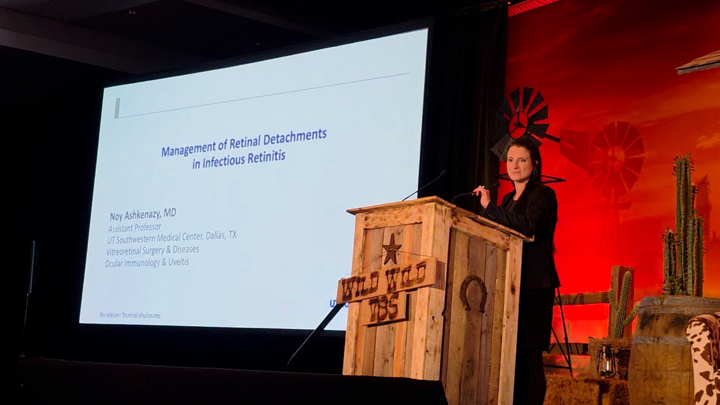

She discussed the case of a 32-year-old woman who presented with a toxoplasmosis lesion associated with a GRT RD with PVR. After PPV with gas, the patient re-detached with PVR, requiring scleral buckle, lensectomy, vitrectomy and silicone oil. Diffuse posterior and anterior fibrotic membranes were encountered, necessitating the use of a microvitreoretinal (MVR) blade for dissection. The patient received systemic antimicrobials as well as intravenous dexamethasone and methotrexate. She underscored the importance of aphakia in cases of anterior PVR and suggested avoiding sub-Tenon’s triamcinolone.
She then showed a diffuse CMV retinitis case in a 28-year-old with ALL who presented with an RD requiring vitrectomy with silicone oil but who then re-detached with PVR, requiring extensive PVR and ILM peeling. She discussed three cases of VZV-associated acute retinal necrosis (ARN) with subsequent retinal detachment. In these cases, she underscored the importance of a scleral buckle to protect the atrophic retina during future vitreous base contraction, avoiding posterior retinotomies given risk of PVR and the use of intraoperative OCT to find micro-breaks if no obvious one is found. She also noted that when the retinitis is diffuse, extensively atrophic retina can necessitate extensive endolaser to ensure retinopexy.
She concluded by discussing timing, noting that it is best to treat viral retinitis aggressively with intravitreal antivirals first (3-6 biweekly injections) before going to the OR to fix an associated RD, as it is important to allow the retina to heal partially from active retinitis. This approach aligns with data indicating a higher risk of re-detachment when surgery is performed in the setting of active inflammation.
Teaching and Learning Advanced Diabetic Vitrectomy – James Rice, MBBCh, MRCOphth, FCOphth(SA), MPH
Dr. James Rice delivered a practical session on how to teach advanced diabetic vitrectomy. He emphasized that developing both technical and mental skills is key—and that this process relies heavily on structured feedback and mentorship.
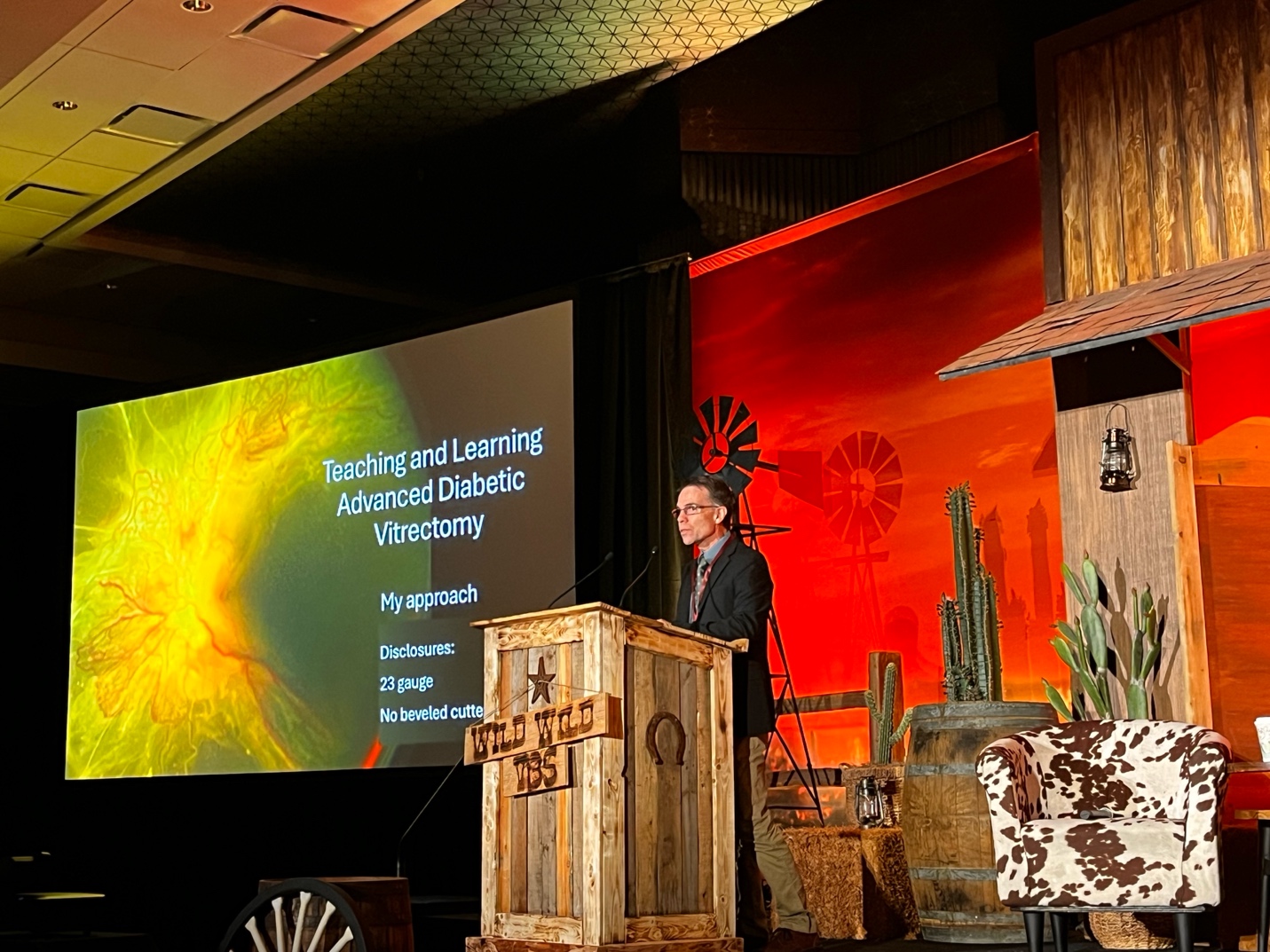

A central part of his talk focused on his step-by-step teaching strategy for managing complex diabetic TRDs: using a posterior-to-anterior (or “inside-out”) approach, find and access the true surgical plane between the hyaloid and the retina early in the case, use the “C-pull” technique to extend hyaloid detachment in a circular movement, reduce the risk of iatrogenic breaks by increasing the distance between the instrument and adhesion points, and use the implosion technique for cutting loose membrane edges. He noted that segmentation of residual membranes is easier with smaller-gauge instruments, and that when membranes are particular tight, creating a space with forceps helps followed by segmentation when there’s a gap. Bimanual surgery is often essential, particularly in combined retinal detachments.
He also discussed the critical role of the educator, especially in providing post-operative feedback. He reviews surgical videos with trainees, uses a progress sheet outlining each surgical step to identify learning gaps, and suggested developing an OSCAR-style scoring sheet specific to diabetic PPV.