Rosalind Ni
Thomas Jefferson University Hospitals, Philadelphia, PA
Vishal Swaminathan
Mayo Clinic, Rochester, MN
The morning of VBS 2025 continued with discussion regarding prominent surgical scientific papers and their impact on clinical practice.
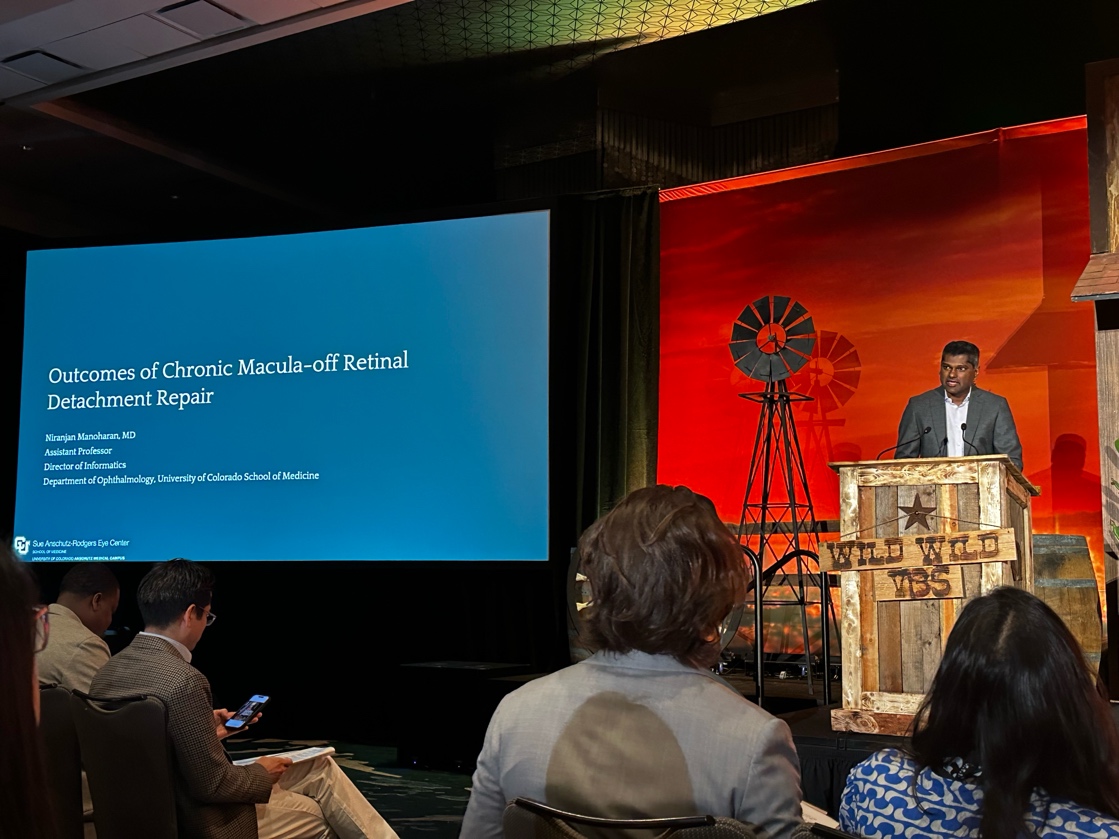
The session began with Dr. Niranjan Manoharan and his talk, “Outcomes of Chronic Retinal Detachment Repair.” He focused upon the potential benefits of operating on end-stage eye disease, particularly diabetic cases or those with chronic rhegmatogenous retinal detachments (RRDs). His study focused specifically on chronic RRDs associated with a posterior vitreous detachment (PVD), which often present with long-standing bullous detachments, chronic macula-off status, and/or proliferative vitreoretinopathy (PVR). Some papers even argue whether these should be repaired at all. In contrast, chronic non-PVD RRDs (e.g., atrophic holes, shallow detachments with demarcation lines) often have a more favorable prognosis.
In his study, he reviewed 37 patients who underwent RRD repair at his local county hospital, focusing on “chronic” RRDs with >30 days of central vision loss, macula-off status, with presence of a PVD, without traumatic detachments. He found an initial success rate of 54% and a final anatomic success of 95%, with an average final Snellen visual acuity of 20/800. He emphasized how repairing chronic RRDs is worthwhile, even in these challenging cases, and that despite long-standing detachment and PVR, final anatomic success is high with appropriate intervention.
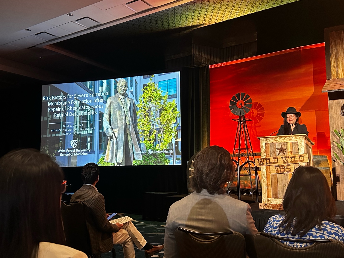

Next, Dr. Sally S. Ong discussed her topic of “Risk Factors for Severe ERM Formation after Repair of RRDs.”
She details the clinical significance of severe epiretinal membrane (ERM) formation and impact on anatomic and functional visual prognosis for patients. Thus, she studied the important question of identifying relevant risk factors to consider for formation of severe ERM after repair of rhegmatogenous retinal detachments (RRDs).
She concluded that greater number of alcohol drinks per week, a history of ocular trauma, and presence of vitreous hemorrhage were associated with more severe ERM formation after RRD repair. She compared her results with that of multiple publications from Japan, Spain, and Boston, all of whom described further considerations such as lack of ILM peeling, location of retinal breaks, mac off status, and visual acuity at baseline.
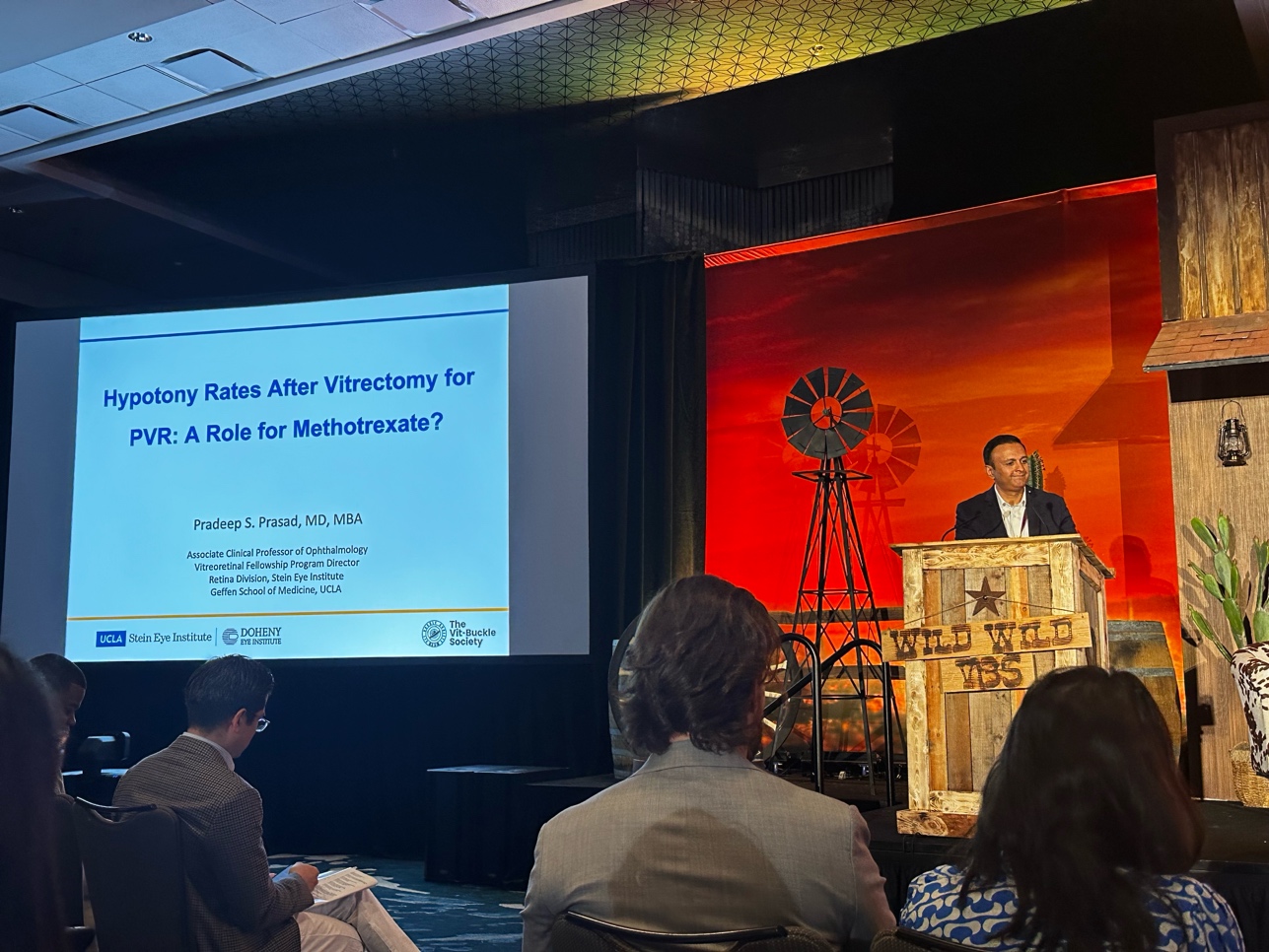

This was followed by an interesting discussion by Dr. Pradeep S. Prasad regarding his topic of “Hypotony After Surgery for PVR: Risk Factors and the Role of Methotrexate.” He discussed how hypotony associated with PVR is a significant unmet clinical challenge. His study focused upon analyzing rates and risk factors for hypotony following tractional PVR repair and investigating whether methotrexate (MTX) has an impact on hypotony rates.
He presented a retrospective study (2018–2024) at his institution in patients with grade C PVR with ≥6 months of follow-up. Patients with preexisting uveitis or glaucoma were excluded and patients receiving at least one MTX injection were placed in the MTX group. The primary outcome was analyzing the incidence of hypotony (IOP <6 mmHg for ≥6 months).
He found that the overall hypotony rate was 13%, which compares favorably with prior studies. Visual acuity was significantly worse in patients who developed hypotony, and risk factors for hypotony included preoperative hypotony, extent of retinal detachment, and retinectomy. Hypotony rates were 4.2% in MTX-treated patients and 17.4% in non-MTX patients. Interestingly, MTX significantly reduced the risk of post-surgical hypotony in PVR cases. The important take-away is that early MTX use may lead to better anatomic and functional outcomes.
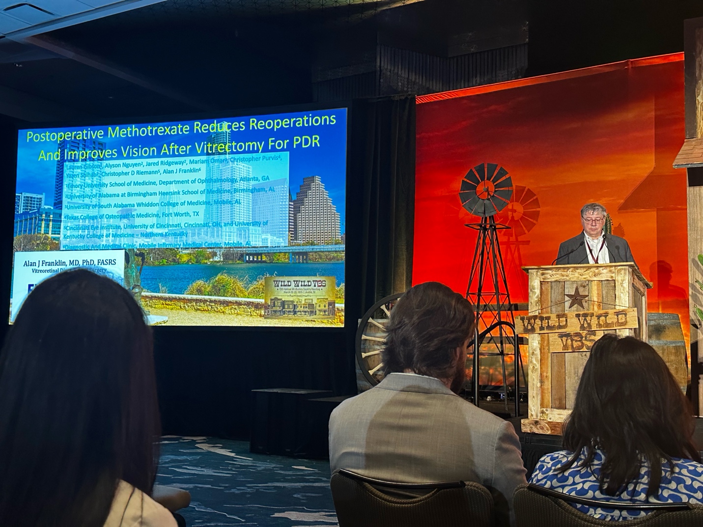

The session continued with Dr. Alan J. Franklin’s presentation on “Methotrexate in Severe PVR & PDR: Impact on Surgical Outcomes.” He discussed the challenges of poor visual prognosis and high reoperation rates when treating patients with retinal detachments associated with severe PVR and proliferative diabetic retinopathy (PDR). The two conditions have similar pathophysiological mechanisms involving TGF-β signaling, nitric oxide synthesis, and hypoxia-driven pathways.
Dr. Franklin presented a retrospective, single-surgeon study evaluating whether intravitreal methotrexate (MTX), which has been studied as a treatment for PVR prevention, improves surgical outcomes in patients with severe PVR, PDR, and trauma-related detachments. Patients each received 3–5 MTX injections (200–400 μg each) and were compared to a control group. The study found that MTX significantly reduced reoperation rates by 57% and increased the likelihood of single-surgery success by 175%. While MTX did not increase the proportion of patients gaining three or more lines of vision, it significantly reduced the rate of three-line vision loss.
Dr. Franklin specifically discussed a notable patient case to illustrate the treatment’s real-world impact—A former football player with bilateral PVR and hand motion vision after an operation in one eye had vision improved to 20/80 after MTX-assisted surgery and was then able to attend an important career honoring event. MTX was well tolerated with minimal safety concerns (~1000 injections performed). Dr. Franklin concluded that MTX is a safe and effective adjunct in managing complex retinal detachments and advocated for further prospective studies and optimization of treatment protocols.
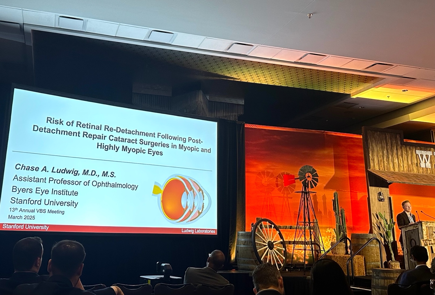

Next, Dr. Chase A. Ludwig presented “Retinal Re-Detachment Risk Following Cataract Surgery in Myopic and Highly Myopic Eyes.” He addressed the common clinical concern of if cataract surgery poses an increased risk for retinal re-detachment in patients with a history of retinal detachment and high myopia, since prior RRD, cataract surgery, and myopia are each independent risk factors for retinal detachment.
This study identified 1,222 individuals who underwent cataract surgery after prior retinal detachment repair from the TriNetX database (+124 million patients). Among these patients, the overall redetachment rate was 8.84%. Redetachment rates appeared to increase with myopia severity, although this trend was not statistically significant. Notably, while single-surgery anatomic success rates were generally favorable, younger patients (ages 18–35) exhibited the highest risk of redetachment at all follow-up intervals, making younger age a significant risk factor for redetachment.
These findings highlight the need for cautious long-term postoperative monitoring, particularly in younger and highly myopic patients. Dr. Ludgwig emphasized the importance of future studies that incorporating axial length and refractive error data for more precise analysis.
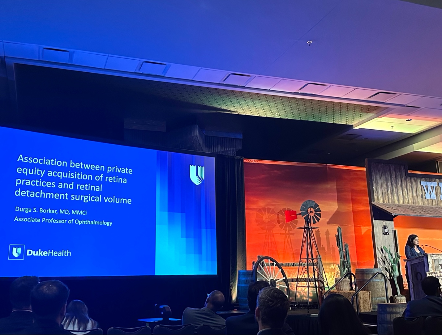

To wrap up this session, Dr. Durga S. Borkar presented on “Association Between Private Equity Acquisition of Retinal Practices and Retinal Detachment Surgical Volume.” Although the incidence of retinal detachment is rising, studies have shown that there is an opportunity cost to performing surgical retinal interventions in the operating room, compared to clinic-based procedures. This study investigated how private equity (PE) acquisitions influenced the volume of retinal detachment surgeries performed.
To compare pre- and post-acquisition surgical volumes, a difference-in-differences analysis compared 535 PE-acquired retina specialists (acquisitions which occurred between 2014-2021) with 1,078 matched controls. The findings revealed a nearly 20% reduction in retinal detachment repair volume in PE-acquired practices post-acquisition. Additionally, there was a significant decline in surgeries for patients with hypertension (5%) and diabetes (10%), suggesting a potential shift toward operating on healthier patients.
These results raise critical questions about access to care, particularly for vulnerable populations, and how PE ownership may be influencing patient selection for surgery. Dr. Borkar emphasized the need for further research into geographic disparities, and long-term outcomes for vulnerable patient populations, such as Medicare and Medicaid patients.