Raziyeh Mahmoudzadeh, MD
Virginia Commenwealth University
The age-related macular degeneration (AMD) session on Thursday September 11th was moderated by Dr. Deeba Hussain and Dr. Thomas Wubben. Here we summarize the exciting talks in this dynamic session.
- The Role of Myeloid Cell Heterogeneity During Spontaneous Choroidal Neovascularization in Vldlr Knockout Mice
Jeremy Lavine, MD, PhD
Chicago, IL
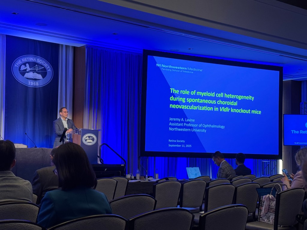
Dr. Lavine opened the AMD session discussing the role of myeloid cells in choroidal neovascularization (CNV), focusing on insights from the Vldlr^-/- mouse model.
Macrophages have long been observed in surgically excised CNV membranes and are known to produce VEGF, promoting angiogenesis in human disease and in laser-induced CNV models. However, the specific myeloid cell subtypes necessary for spontaneous CNV remain unclear.
To investigate this, his team systematically depleted different myeloid populations. Classical and non-classical monocytes were targeted using Ccr2^-/- and Nr4a1^-/- mice, respectively. Neither manipulation affected macrophage counts, CNV lesion number, or total CNV area, suggesting that monocytes and monocyte-derived macrophages are dispensable in this model.
Next, microglia were depleted using two genetic strategies: Vldlr^-/-Tmem119CreER/+Rosa26DTR/+ mice and Vldlr^-/-Cx3cr1CreER Csf1riDTR mice treated with a single low dose of tamoxifen to selectively target microglia. Both approaches reduced macrophage counts and lesion number, but total CNV area remained unchanged, indicating that microglia likely function as initiators of CNV lesions.
The most pronounced effects were observed when both microglia and choroidal macrophages were ablated, either genetically with multiple tamoxifen doses or pharmacologically via CSF1R antagonism. This resulted in dramatic reductions in macrophage counts, CNV lesion number, and total lesion area, suggesting that choroidal macrophages are crucial for CNV growth and/or maintenance.
In summary, monocytes and their derivatives are largely dispensable in Vldlr^-/- mice. Microglia appear to seed CNV lesion formation, while choroidal macrophages sustain lesion growth. These findings highlight the translational potential of systemically targeting choroidal macrophages as a therapeutic strategy for CNV in AMD.
- Targeting Photoreceptor Metabolic Adaptations in Age-Related Macular Degeneration by Modulating Pyruvate Kinase M2 Slows Progression of Disease
Thomas Wubben, MD, PhD
Ann Arbor, MI
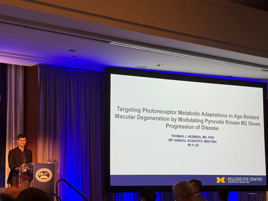

The second talk in the AMD session focused on the role of photoreceptor metabolism in driving AMD pathogenesis, with particular attention to the mTORC1-PKM2 signaling axis. Dr. Wubben began by highlighting that, while much AMD research has focused on complement-mediated inflammation or RPE biology, emerging preclinical and clinical evidence underscores photoreceptors as key players in both early and late AMD. Dysfunction and death of photoreceptors are central to vision loss, and their altered metabolism contributes to AMD-like pathologies.
Using a TSC1 knockout mouse model with hyperactive mTORC1 signaling specifically in photoreceptors, the team recapitulated early and late AMD features. Importantly, they observed that photoreceptors in this model, like those in AMD patients, displayed increased levels of PKM2 in its low-activity dimeric state, which correlated with decreased overall pyruvate kinase activity and metabolic dysregulation.
To test whether correcting this metabolic defect could alter disease progression, the investigators replaced PKM2 with the constitutively active PKM1 isoform in photoreceptors. This genetic intervention increased retinal pyruvate kinase activity, reduced mTORC1 hyperactivation, lowered HIF-1α levels, and normalized the expression of downstream pro-angiogenic and inflammatory mediators, including VEGFA and IL1B. Remarkably, this approach improved RPE mitochondrial function, despite no direct genetic manipulation of RPE cells, and significantly reduced both dry and wet AMD-like pathologies, including subretinal neovascularization.
Pharmacologic activation of PKM2 similarly reproduced these protective effects, demonstrating translational potential. The findings support a model in which photoreceptor metabolic adaptations contribute to AMD pathogenesis and identify PKM2 as a photoreceptor-specific therapeutic target capable of mitigating disease progression.
- Long-Term “Real World” Western-Style Diet and Mediterranean-Type Diet Influence Choroidal Neovascularization and Subretinal Microglia/ Macrophage Infiltration via Gut Microbiome Modulation
Dimitra Skondra, MD, PhD
Chicago, IL
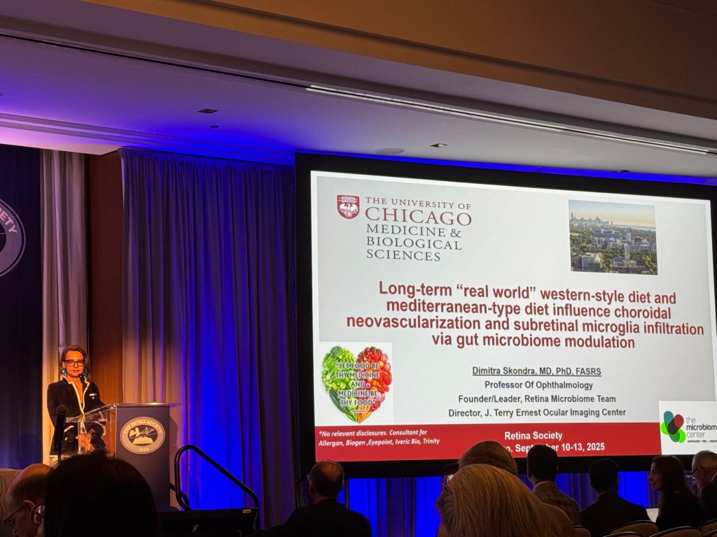

Dr. Skondra began by highlighting the emerging role of diet and the gut-retina axis in influencing susceptibility to neovascular AMD (nAMD), focusing on how long-term dietary habits shape subretinal angiogenesis and immune cell infiltration. She noted that while AMD is common, progression to advanced disease varies among individuals, and upstream factors—such as diet and microbiome composition—may contribute to this differential susceptibility.
Epidemiological evidence has suggested that Western-style diets, high in fats and sugars, increase AMD risk, whereas Mediterranean-type diets rich in fish and plant-based foods appear protective. However, causality and underlying mechanisms have remained unclear. Dr. Skondra highlighted that the gut microbiome likely mediates these diet effects, with “healthy” diets promoting anti-inflammatory microbial metabolites (e.g., short-chain fatty acids) and Western diets favoring pro-inflammatory metabolites (e.g., phenols and palmitate).
To investigate these relationships, the team fed C57BL/6 mice either a Western-style diet, a Mediterranean-type diet, or a standard diet for six months—representing long-term human dietary habits—followed by laser-induced CNV. Choroidal flatmounts and IBA1 staining revealed that Mediterranean-fed mice had significantly smaller CNV lesions and reduced subretinal microglia/macrophage infiltration compared to Western-fed and standard diet groups. Notably, even a standard “ultra-healthy” mouse diet did not outperform the Mediterranean diet in limiting CNV and immune cell infiltration, highlighting the translational relevance of real-world dietary patterns.
Microbiome analyses demonstrated distinct gut microbial fingerprints for each diet. Western-fed mice showed enrichment of inflammatory taxa such as Lactococcus, Streptococcus, and Turicibacter, along with elevated pro-inflammatory metabolites. In contrast, Mediterranean-fed mice had increased abundance of taxa like Ruminococcaceae, Lachnoclostridium, and Dubosiella, which were associated with higher levels of anti-inflammatory metabolites (propionate, butyrate, valerate, cysteine, and glycine). Computational analyses confirmed that diet-driven changes in microbial pathways corresponded to these metabolite profiles. Fecal microbiota transplantation experiments further supported a causal link, as phenotypes associated with the donor diet were recapitulated in recipient mice.
Dr. Skondra concluded by emphasizing that long-term dietary habits can modulate CNV susceptibility through the gut microbiome, shaping subretinal immune responses and angiogenesis. Mediterranean-type diets suppress pro-angiogenic and pro-inflammatory pathways, suggesting dietary modulation of gut microbiota as a promising, non-invasive strategy to reduce risk and progression of nAMD.
- Metabolic Profiling of Normal Human Retinal and Choroidal Tissue and Correlations with Vitreous Humor and Serum
Deeba Husain, MD
Boston, MA
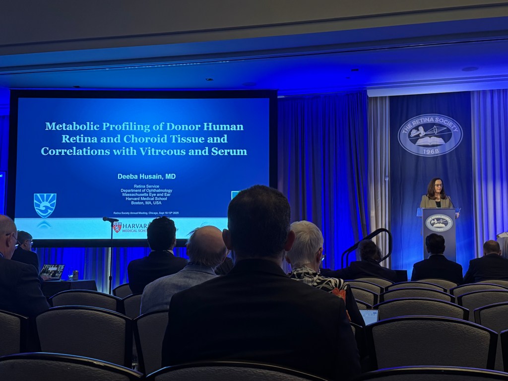

Dr. Husain began by highlighting that metabolomics—the study of small molecules under 1 kDa—reflects the end products of genetic and environmental influences, making it closely linked to disease phenotypes. While previous work in her group demonstrated distinct metabolomic profiles in the biofluids of AMD patients, the relationship between circulating metabolites and ocular tissue metabolites remained unclear.
To address this, the study characterized the metabolomic profiles of normal human donor retina and choroid and assessed correlations with donor serum and vitreous humor. Fourteen donor eyes from individuals over 50 years old with no known retinal disease were collected within six hours post-mortem. The anterior segment and corneal buttons were removed, vitreous was collected, and 6-mm macular and peripheral punches were harvested. Neurosensory retina and RPE/choroid were separated, producing four tissue samples per eye. Serum samples from the same donors were also collected. All samples were snap-frozen and analyzed via non-targeted mass spectrometry at Metabolon Inc., with tissue, vitreous, and serum analyzed together to minimize batch effects.
The analysis identified 659 metabolites in tissue, 419 in vitreous humor, and 958 in serum. Lipids represented the largest class in tissue (53%), followed by amino acids (21%), nucleotides (7%), carbohydrates (6%), cofactors/vitamins (6%), xenobiotics (3%), peptides (2%), and energy-related metabolites (2%). Metabolic profiles were highly consistent between macular and peripheral regions, as well as between neurosensory retina and RPE/choroid, with 70% of metabolites shared across tissue types. Unique metabolites were enriched in amino acids and cofactors for the neurosensory retina and in lipids for the RPE.
Comparisons with biofluids disclosed substantial overlap: 513 metabolites were shared between tissue and serum, predominantly lipids and phospholipids, while 297 were shared with vitreous, mainly amino acids. Dr. Husain emphasized that these findings support the use of circulating metabolites as potential biomarkers of retinal health and disease and provide a foundation for future translational studies linking systemic and ocular metabolism.
- Plasma Metabolomic Profiles Associated with Five-Year Progression of Age-Related Macular Degeneration
Ines Lains, MD, PhD
Boston, MA

Dr. Lains presented longitudinal metabolomic profiling to better understand AMD progression. While cross-sectional studies have highlighted the relevance of metabolomics in AMD, longitudinal analyses over extended periods have been limited. This study aimed to evaluate whether baseline plasma metabolites are associated with AMD progression over five years.
The study was a prospective longitudinal analysis including patients over 50 years old from Boston, USA, and Coimbra, Portugal. Only eyes with non-advanced AMD at baseline were included, and patients with diabetes were excluded to avoid confounding effects on the metabolome. Participants underwent color fundus photography (CFP) and optical coherence tomography (OCT) at baseline and after five years. Fasting plasma samples were collected at both time points. Eyes were classified as “progressed” if their AMD stage (AREDS classification) advanced over five years.
Methodologically, the study accounted for correlated data from two eyes of the same patient. Each cohort was analyzed separately, followed by a meta-analysis to combine results, which is what Dr. Lains presented. OCT images were graded by two independent graders for features such as hyperreflective foci, subretinal drusenoid deposits, and atrophy. Plasma metabolomic profiling was performed using mass spectrometry (Metabolon, Inc.).
The study included 361 eyes (131 US, 230 Portugal), with 81 eyes (22.4%) showing progression at five years. Based on CFP analysis, two baseline metabolites—lactate and a branched-chain lipid—were significantly associated with progression. OCT analyses revealed that 37 baseline metabolites were associated with development of hyperreflective foci, the majority being glycerophospholipids (glycerophosphatidylcholines and glycerophosphatidylmethanolines). Thirty-two metabolites were associated with the development of atrophy, including sphingolipids and amino acids, while three lipids were linked to the formation of subretinal drusenoid deposits.
Dr. Lains emphasized that these findings suggest baseline plasma metabolomic profiles, particularly lipid metabolites, reflect susceptibility to AMD progression and OCT features over five years. This study provides novel insights into AMD pathophysiology and highlights potential biomarkers for early identification of high-risk patients, advancing our understanding of the disease’s natural history.
- Association Between Systemic Complement Levels and Multimodal Imaging Biomarkers in Intermediate Age-Related Macular Degeneration
Talisa Eiko de Carlo Forest, MD
Aurora, CO
Dr. de Carlo Forest closed the session by addressing the interplay between systemic complement dysregulation and imaging biomarkers in intermediate AMD (iAMD). She emphasized that both systemic and local immune activation, particularly through the complement system, have long been implicated in AMD pathogenesis, which is why many emerging therapies target complement pathways. At the same time, select imaging biomarkers are recognized as strong predictors of disease progression. This study brought those two dimensions together by asking whether high-risk imaging features are associated with systemic complement activity.
The analysis drew from the University of Colorado AMD Registry, a longitudinal cohort of more than 1,600 patients. For this cross-sectional study, approximately 436 patients with iAMD who had both multimodal imaging and plasma samples available were included. Imaging was graded by two trained readers for the presence of drusen, drusenoid PED, calcified drusen, hyperreflective foci, and geographic atrophy. Systemic complement factors and activation ratios across classical, alternative, and terminal pathways were measured using ELISA assays. Analyses were performed with nonparametric tests and multivariate logistic regression adjusted for age, with false discovery rate correction.
Key findings showed that moderate-risk biomarkers—including soft drusen, drusenoid PED, and calcified drusen—were consistently associated with upregulation of complement markers across multiple pathways. By contrast, higher-risk features such as hyperreflective foci and geographic atrophy were linked to a relative downregulation of complement activity. Dr. de Carlo Forest suggested this pattern reflects dynamic changes in systemic complement regulation: an upregulation during mid-stage disease, followed by relative downregulation as disease advances.
These results reinforce the role of complement dysregulation in iAMD, while also raising important questions about when in the disease course complement-targeted therapy may be most effective. Importantly, the study also highlighted that not only the alternative pathway but also the classical and terminal pathways appear to contribute meaningfully. Future work will focus on longitudinal analyses to better define how systemic complement levels evolve over time and how they interact with imaging and genetics.