Giulia Corradetti, MD
Doheny Eye Institute, UCLA
At the FLORetina-ICOOR meeting, on December 1, 2023, the OCT-Angiography Course was presented in the Ellisse Room and moderated by Drs. Daniela Bacherini, Marion Munk, and Maria Cristina Savastano.
Dr. Maria Cristina Savastano from the University Cattolica Sacro Cuore, Rome, presented the first talk of the session entitled “Hyperreflectivity and hyporeflectivity in OCTA: new biomarkers.” Dr. Savastano introduced her presentation by highlighting that hyporeflective and hyperreflective elements in OCTA imaging can be located at any level in the retina and choroid, thus she presented a series of cases displaying the related multimodal imaging, including the OCTA characteristic imaging.
A broad variety of diseases were discussed: perifoveal exudative vascular anomalous complex (PEVAC), acute retinal necrosis (ARN), cilio-retinal artery occlusion in Horton disease, acute retinal venous occlusion (RVO) secondary to epiretinal membrane removal, paracentral acute middle maculopathy (PAMM) and acute macular neuroretinopathy (AMN), diabetic retinopathy, macular neovascularization, non-neovascular AMD, late atrophic AMD, multiple evanescent white dot syndrome (MEDWS), full thickness macular hole (FTMH).
Dr. Savastano concluded by highlighting the importance of multimodal imaging in the diagnosis and management of retinal diseases, allowing for a more precise interpretation of hyperreflective and hyporeflective OCTA signals.

Dr. Marion Munk (above) then presented the second talk of the session entitled “Cultivating Nomenclature in Optical Coherence Tomography Angiography – An Update on Standardization Efforts.” She highlighted that there is no consensus to date on the OCTA nomenclature and a number of contradictory terms have been used to define and describe OCTA findings in the literature. Establishing a consensus nomenclature for the description, interpretation, and reporting of OCTA images would be pivotal in enhancing the standardization of the quantitative computational analyses.
Dr. Munk illustrated the consensus results of surveys distributed among the members of the International Imaging Retinal Society (IntRIS). The voting participants were selected from multiple countries and were invited based on their expertise in OCTA imaging. The experts agreed 1) to implement the description of the OCTA signal with adjectives such as “increase, decrease, normal or abnormal OCTA signal”and 2) to use descriptive terms to describe the scan type, to define the scan pattern, and precisely report the location of the OCTA slab (e.g. deep capillary plexus versus superficial capillary plexus).
Based on the results of these surveys, a new framework for reporting OCTA will be presented to achieve a greater standardization of the interpretations and the description of OCTA findings in clinical settings, clinical trials, and publications.
Dr. Marco Rispoli presented a case series of OCTA in acute idiopathic maculopathy, highlighting the importance of a multimodal approach. Dr. Rispoli illustrated the proposed disease staging of acute idiopathic maculopathy based on multimodal imaging, in which 4 stages were detected based on the fundus autofluorescence characteristics. He remarked on the importance of recognizing typical patterns of acute idiopathic maculopathy, that could optimize clinical diagnosis and patient counseling.
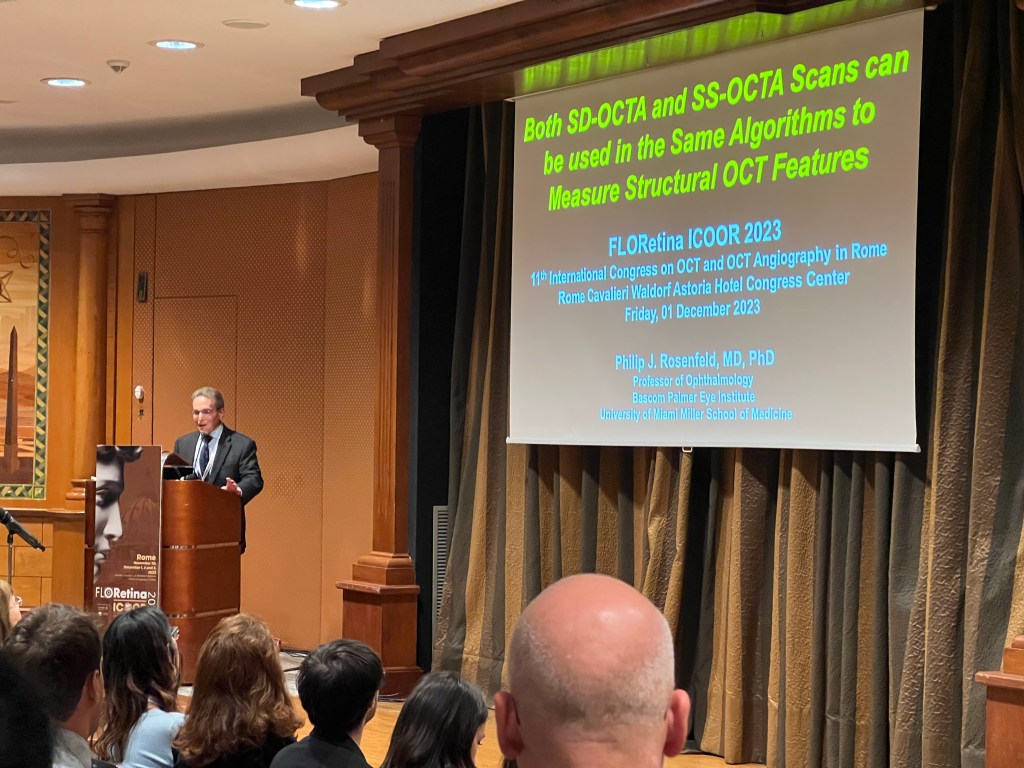

Dr. Philip J. Rosenfeld (above) from Bascom Palmer Institute, Miami, presented a talk commenting on the application of algorithms to measure structural OCT features on both SD-OCTA and SS-OCTA devices. While SS-OCTA scans can be used to identify many structural biomarkers, SS-OCTA devices are not widely available yet. Dr. Rosenfeld illustrated that SS-OCTA algorithms are compatible with SD-OCTA scans to measure drusen volume and drusen area, and hypertransmission defects (HTDs). Although SS-OCTA instruments provide a more accurate segmentation, there are no clinically significant differences in the measurements between the two types of devices. Dr. Rosenfeld concluded that SS- and SD-OCTA scans both contain important structural information that can be processed using the same algorithms.
Dr. Jia Yali presented a lecture entitled “Active and inactive microaneurysms identified and characterized by OCTA” by illustrating examples of perfused and non-perfused microaneurysms identified and characterized on OCT and OCTA scans. Dr. Yali highlighted that microaneurysms have distinct, oval-shaped, hyperreflective walls on structural OCT with inconsistent flow in the lumen with OCTA.
He presented the approach his team adopted for classification of microaneurysms on OCTA. First, in the presence of the microaneurysms, the state of perfusion or non-perfusion was assessed, based on the presence of flow signal within the lumen of the microaneurysms from cross-sectional OCTA.
En face OCTA image evaluation represented the next step: perfused microaneurysms showed a well-defined vascular outpouching on en face OCTA, whereas the partially perfused showed a weaker signal.
By comparing the detection of microaneurysms with OCT, OCTA, and FA, 6 different scenarios were described. He highlighted that microaneurysms are best detected on volume-averaged high-definition OCTA, and most microaneurysms on OCT/OCTA are perfused and associated with retinal fluid.
In conclusion, the OCT/OCTA evaluation of the perfusion state of microaneurysms may be important in planning the treatment of diabetic macular edema.
Dr. Hormel Trista (below) then presented a talk on “High-resolution wide field single shot panretinal OCTA” by highlighting that one of the main limitations of current OCTA is the limited field of view. Dr. Hormel presented a swept-source OCTA prototype using a 500 kHz high-speed laser, which can generate 75-degree field of view without need for mydriasis in a single acquisition. A series of examples of retinal diseases captured on the OCTA prototype were shown.

Dr. Cohen Yves Salomon introduced his talk with the question “Is OCTA really useful in daily practice?” and presented the limitations and advantages of OCTA imaging in different retinal diseases.
Dr. Amani Fawzi (below) presented a lecture entitled “Retinal capillary perfusion: do OCTA and ultrawide-field FA correlate?” She illustrated the quantitative definition of pathological non-perfusion in diabetic retinopathy and described published algorithms to assess non-perfused areas. She talked about the correlation between non-perfusion parameters and ultrawide-field fluorescein angiography in the context of diabetic retinopathy. Dr. Fawzi concluded that non-perfused areas as quantified on OCTA correlated well with ultrawide-field FA parameters. Therefore, macular OCTA metrics, especially geometric perfusion density at the level of the deep capillary plexus has the potential to describe the overall ischemic state of the retina, including the periphery.

Dr. Daniela Bacherini delivered the last presentation of the session, entitled “OCTA in Emergencies” illustrating a series of patients with central retinal arterial occlusion who presented to the emergency department, and underwent ocular massage. She showed the beneficial effects reported on OCTA scans. Dr. Bacherini highlighted the applications and importance of OCTA, as an innovative non-invasive retinal imaging tool in clinical and emergency settings.